Virtual nanoscopy
Posted by Eva Amsen, on 21 August 2012
Let’s take a very close look at the inside of a fish!
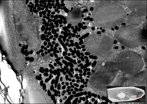
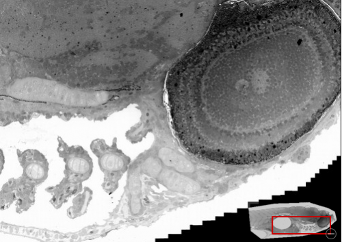
A recent paper in the Journal of Cell Biology describes a technique for generating large, composite, images from electron microscopy data. Frank Faas, Raimond Ravelli, and colleagues at the Leiden University Medical Center developed a method to computationally collect and align EM images. In the data viewer accompanying the paper, they show a large section of a zebrafish embryo, 5 days post-fertilisation, which is comprised of 26,434 individual images! The total size of the composite image is 921,600 pixels by 380,928 pixels. (For reference, the screenshots below are 500 pixels wide.)
In these screenshots you can see the overall fish in the bottom right, with the red area indicating where the larger, detailed, view is located. These are two different magnifications of the (edge of the) eye. In the bottom image you can also see the edge of the composite image, and appreciate how many individual images there are, and how well they connect.


To find out more about the method, and its practical applications, read the full paper and editorial at JCB.
![]() Faas FG, Avramut MC, M van den Berg B, Mommaas AM, Koster AJ, & Ravelli RB (2012). Virtual nanoscopy: Generation of ultra-large high resolution electron microscopy maps. The Journal of cell biology, 198 (3), 457-69 PMID: 22869601
Faas FG, Avramut MC, M van den Berg B, Mommaas AM, Koster AJ, & Ravelli RB (2012). Virtual nanoscopy: Generation of ultra-large high resolution electron microscopy maps. The Journal of cell biology, 198 (3), 457-69 PMID: 22869601


 (2 votes)
Thanks Eva – very nice!