The 3D atlas of human development
Posted by 3DEmbryo, on 28 November 2016
“It’s fair to say that we currently know more about the moon than about our own embryonic development. The current textbooks all show the same kind of images based on a few embryonic specimens from the 1930s. Some of those are not even human embryos. New version of these books keep using those images, often without reference, only updating new molecular information”
This leads to the situation that the origin of the knowledge on human embryology became forgotten and that some of the knowledge was based on chicken, axolotl and mouse specimens. Moreover, the complex morphological changes during development cannot be well illustrated on the flat two-dimensional pages of text books.
To remedy this situation we proposed to use the sections of the embryos available in the Carnegie Collection to generate a comprehensive series of interactive 3D reconstructions of human embryonic development; a proposal that was based on the availability of 3D reconstruction and visualisation software.
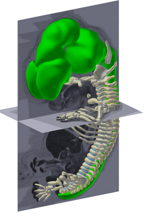
This project, initiated by Professor Antoon Moorman, with dedicated financial support of the Academic Medical Center in Amsterdam, the Netherlands, was carried out by Bernadette de Bakker. The educational angle made that the project was ‘by students – for students’. De Bakker obtained a duplicate series of images of stages 7 to 23 (15 – 60 days of development) from the Carnegie Collection in Washington DC. After alignment in a 3D reconstruction program, a total of 75 medical students manually annotated every structure and organ in each of the about 15,000 images as part of their scientific internship; an effort that took more than 45,000 hours.
Another 15 students, this time from the game development faculty, then ‘modelled’ the resulting 3D reconstructions to reduce the complexity without losing biological detail. Their results, turned into 3D-PDF files, thus represent the actual morphology of the series of embryos in a format that can be easily interactively handled with the commonly used PDF reader. This was all supervised and supported by the expert embryologists of the department.
“The 3D Atlas and Database of Human Embryology is the first to present such a large amount of data based on such a large number of human embryos. With this atlas and database we are able to quantify very precisely embryonic growth and the growth and position of specific organs”
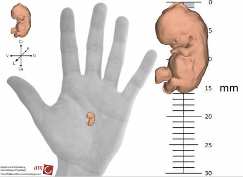
The 3D atlas, that contains morphological reconstructions of 14 stages, is accompanied by a database with quantitative data based on 17 duplicate stages of reconstructed embryos. This database shows the total volume of the embryo grows exponentially with a constant volume increase of 25% per day. However, different organs grow with different growth rates, depending on their function during development. The project team established a tool to determine the position of organs relative to the developing vertebral column of the embryo. Application of this tool can help to solve ambiguities with respect to the relation between organs. The third series of data relates the chronology of human development to that of the mouse and the chicken. These data show that each species runs its own embryological script.
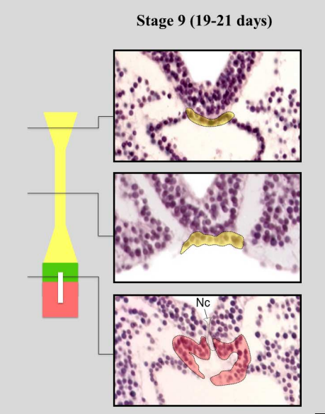
Already during the construction of the atlas, discrepancies between the observed embryology and the embryology textbooks became apparent. The textbook descriptions of the development of the venous system and the notochord clearly show influence of knowledge dissipating from studies on lower vertebrates. Descriptions that could not be corroborated in the current reconstructions based on the real human substrate. The interactive 3D atlas, and the source images that are also available from the website, allow every user, be it student or biomedical specialist, to independently verify those discrepancies and form their own opinion.
“The atlas is useful because to study how congenital malformations appear it is important to know how normal human embryonic development occurs. Experimental embryologists will know how the changes in their experimental animals relate to the human situation. Scientists who study how toxic substances can affect embryonic development, are helped to refine the chick and mice models they work with. Moreover, also gynaecologists can use this atlas to show pregnant women how their child develops.”
Contributors: Bernadette de Bakker, Jan M Ruijter and Antoon Moorman
The 3D atlas of human embryology is available here:
The Research Article in Science reporting the atlas is available here, Open Access:
http://science.sciencemag.org/content/354/6315/aag0053
 (2 votes)
One thought on “The 3D atlas of human development”
Leave a Reply
Get involved
Create an account or log in to post your story on the Node.
Sign up for emails
Subscribe to our mailing lists.
Fetal development to 3 weeks and 8 weeks