A New Way To Look At Human Development
Posted by samuel.malone, on 29 March 2017
Throughout history, the desire of scientists to understand physiology and disease by thoroughly studying anatomical features, has always faced an intractable limitation: they cannot simply see through the tissue! Dissection has therefore been the modus operandi of anatomists: from Galen’s pioneering studies, to modern day biologists who routinely section tissues to label structures for microscopic analysis.
Whilst these methods have informed a wealth of knowledge linking anatomical form to function, they are inherently flawed due to a 3-Dimensional appreciation of structures being lost. This has been especially problematic for the study of human development, where structures are continually evolving, and therefore a precise visualisation has been impossible to achieve through traditional methods and anatomical atlases. Coupled with the difficulties in tissue access, our understanding of human development has progressed perhaps the slowest of any biological process since the 1930’s; whilst in some cases observations of lower vertebrates have subsequently been erroneously applied to humans.
“Birth defects of structural or functional origin currently affect more than 3% of births”
Without a good understanding of physiological development, we lack the fundamental knowledge required for clinicians and researchers to tackle a healthcare issue that inflicts a severe healthcare and emotional burden. Recent advances in non-invasive, in vivo imaging techniques, have shown great promise in detecting congenital abnormalities as well as providing information on gross topological features of fetal development; however, they lack sufficient resolution in order to inform developmental biologists of currently unknown features of organogenesis. This has recently been most strikingly highlighted by the surge in Zika virus infections and reports of its detrimental effects on cephalic development.
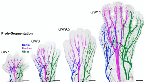
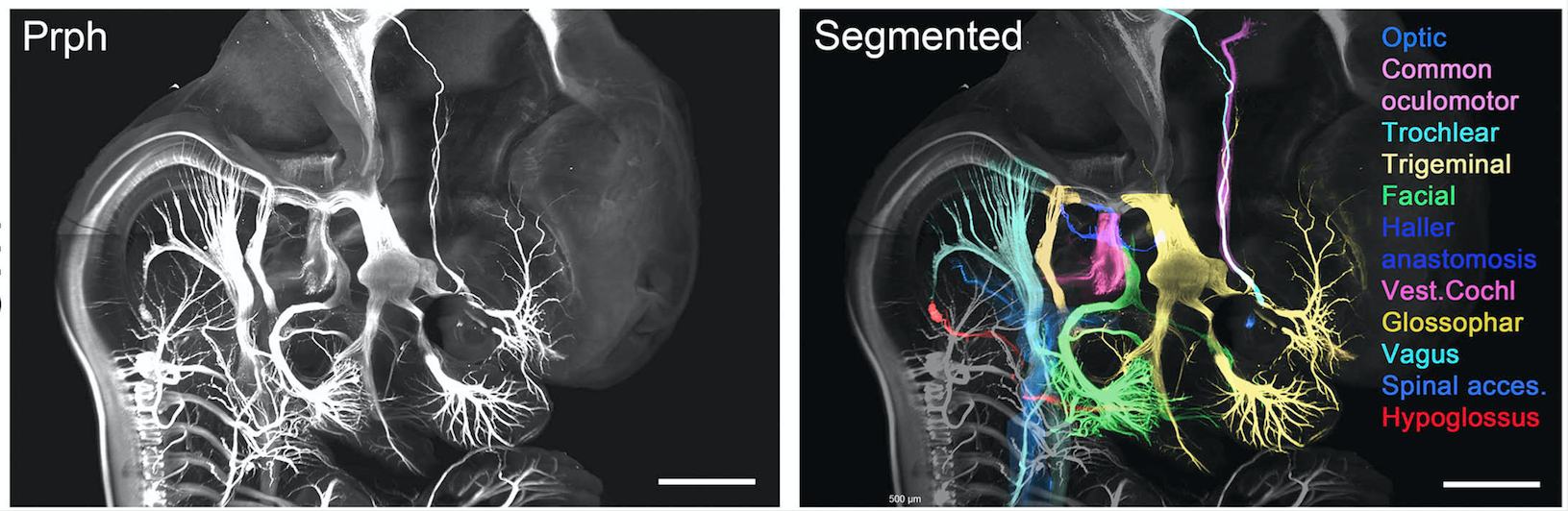
In order to begin rapidly addressing the gaping holes in our knowledge, we have begun to form a cutting edge research consortium of developmental biologists, to build a detailed human cell atlas. Here we have expanded the use of clearing techniques to human tissues and immunolabeled with over 40 antibodies, intact human embryos during the first trimester of gestation (from 6 to 14 gestational weeks).
In recent years, several techniques have been developed for tissue clearing, in which whole organs are rendered macromolecule permeable and optically transparent. Among tissue-clearing techniques, the process called 3D imaging of solvent-cleared organs, or 3DISCO has been proved to be a simple, robust and inexpensive method for 3D analysis of immunolabeled transparent organs in embryonic and postnatal mice.
Currently organised along seven organ systems, the project aims to expand and evolve as more data is added. Current data in the atlas examines molecular organogenesis based on over 40 samples comprising 1,500,000 optical sections – making it the most complete 3D analysis of early human development currently available.
Constructed by imaging intact tissues and whole embryos, the atlas will be an invaluable tool for researchers with the ability to explore cell distributions, count proliferating cells in each organ etc. whilst also being useful for didactic purposes, with the ability to 3D print models to inform health science teaching programs.
This has long been the dream of developmental biologists, which has finally been realised by the use of organic solvent based clearing techniques (3DISCO/iDISCO) combined with light sheet imaging. Together, these powerful approaches allow inexpensive labelling of any cell population of all developing organ systems during development and imaging at cellular resolution whilst fully maintaining structural relationships. To further highlight the robustness and power of this technique to analyse human development, it should be noted that these data were generated in just over one year by a small team of researchers.
And this is just the beginning, as the online resources are open access – available for researchers all over the world to analyse and contribute to, along with new data we will acquire. The goal of the project is to make a continuously updated 3D molecular reference atlas of human cells during development, paramount to a better understanding of human development during health and disease.
The online video series of immunolabeled tissues along with the original data sets is available at https://transparent-human-embryo.com/ . The 3D database was developed with Keen eye technologies with support from the “fondation voir et entrendre”
The original research paper featured in Cell is available here: Tridimensional Visualization and Analysis of Early Human Development
Consortium spearheaded by Dr. Alain Chedotal of the Institut de la Vision, Paris, France & Dr. Paolo Giacobini, University of Lille, France
By Samuel A. Malone
 (3 votes)
Get involved
Create an account or log in to post your story on the Node.
Sign up for emails
Subscribe to our mailing lists.