Who won the 2024 Development cover image of the year?
Posted by the Node, on 15 January 2025
Over the last few weeks, we asked you to vote for your favourite 2024 Development cover image. Thank you to everyone who voted. Now that the poll is closed, let’s reveal the results!
*Drumroll*
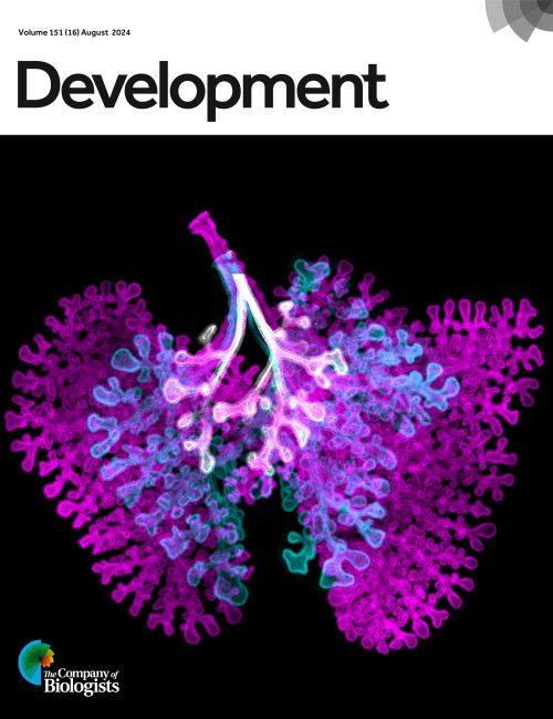
The 2024 Development cover of the year is the image of a superimposition of three stages of embryonic mouse lungs! Congratulations to Paramore et al.!
Browse the full 2024 issue collection, including our Special Issue: Uncovering Developmental Diversity.
Winner

A superimposition of three stages of embryonic mouse lungs (E12 in white, E13 in cyan and E14 in magenta) demonstrating changes that can be observed over a 3-day period. The pulmonary mesenchyme regulates the lengthening and widening of airways via the protein Vangl2, revealing a previously unreported role for this tissue compartment in the shaping of the airway tree. See Research article by Paramore et al.
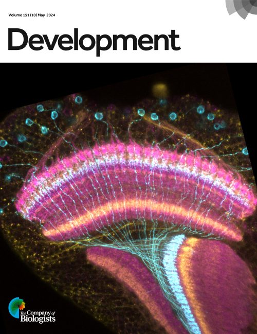
First runner-up

Drosophila optic lobe at 72 hours after puparium formation. Tm9 neurons are labelled with GMR24C08-GAL4 expressing UAS-myristoylated Tomato (cyan), the medulla, lobula and lobula plate neuropils are labeled with anti-N-cadherin (magenta), and specific layers of these neuropils are labeled with anti-connectin (yellow). Image credit: Maria Bustillo. See Research article by Bustillo et al.
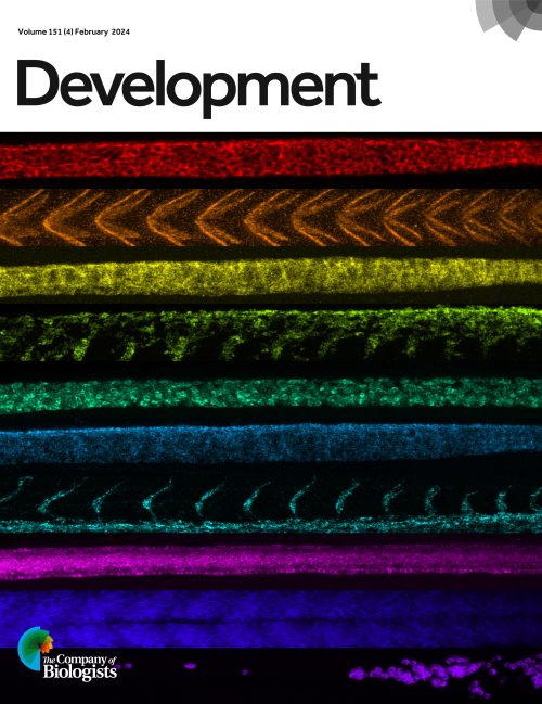
Second runner-up

Collage of RNA expression in the tail of a whole-mount zebrafish embryo composed from the channels of a 10-plex, quantitative, high-resolution RNA fluorescence in situ hybridization experiment performed using spectral imaging with signal amplification based on the mechanism of hybridization chain reaction (HCR). See Research article by Schulte at al.
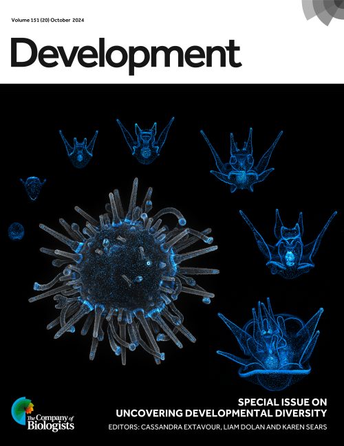
Honourable mention

Development of transgenic Lytechinus pictus, the first transgenic echinoderm lines, expressing cyan fluorescent protein fused to a nuclear marker (histone 2B) driven by a polyubiquitin promoter. Developmental stages expressing the transgene are depicted from blastula (12 h post-fertilisation) through the larval stages, to the competent larva (22 days post-fertilisation), and finally to the juvenile stage at center. The juvenile has an additional membrane stain (grey) for contrast. See Research article by Jackson et al.
Browse the full 2024 issue collection, including our Special Issue: Uncovering Developmental Diversity.
We look forward to seeing more amazing cover images featured in Development in the coming year!


 (No Ratings Yet)