The 2025 Developmental Biology Gordon Research Seminar (GRS) was held on March 29-30 2025. This year, the GRS co-chairs Ana R. Hernandez Rodriguez and Anastasia Repouliou decided to organise an image competition by asking the GRS participants to submit their images and vote for their favourite.
“I have always believed that one of the great strengths of developmental biology is its remarkable visual appeal. The Developmental Biology GRC was a wonderfully diverse meeting with a variety of established and emerging model organisms, and we wanted to provide a platform for researchers to share captivating images of their work. These images were displayed throughout the week during breaks and poster sessions, creating an inspiring backdrop for cutting edge scientific discussions and highlighting the diversity and creativity within our community.”
Ana Hernandez (Co-Chair GRS Developmental Biology 2025)
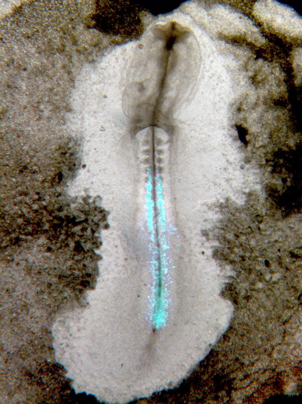
Among the great selection of images, Paul Bump’s “Life as a drop in the ocean” won the most votes – congratulations!
You can browse through all the great entries to the competition in the image gallery below.
Browse through the image gallery (click to expand)
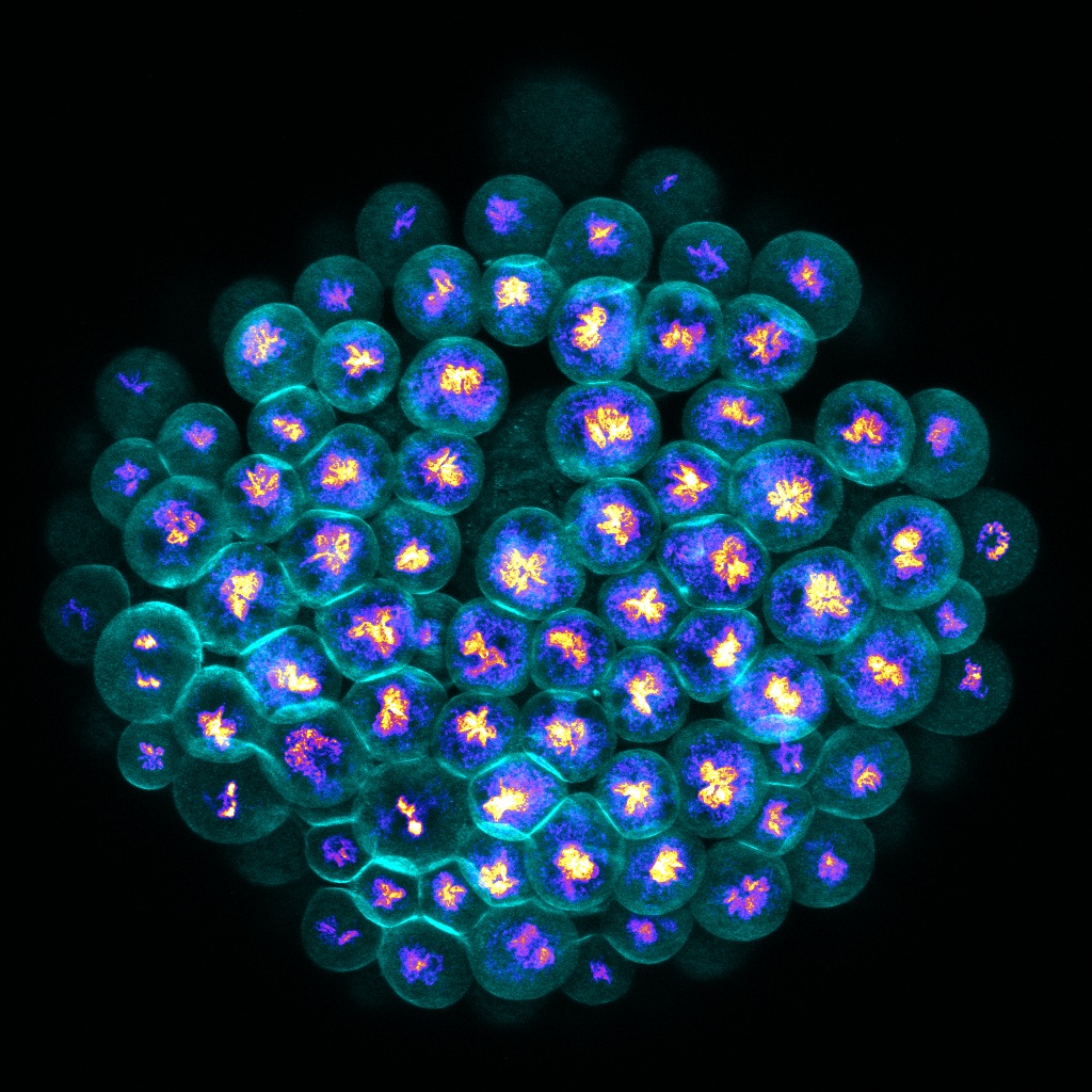
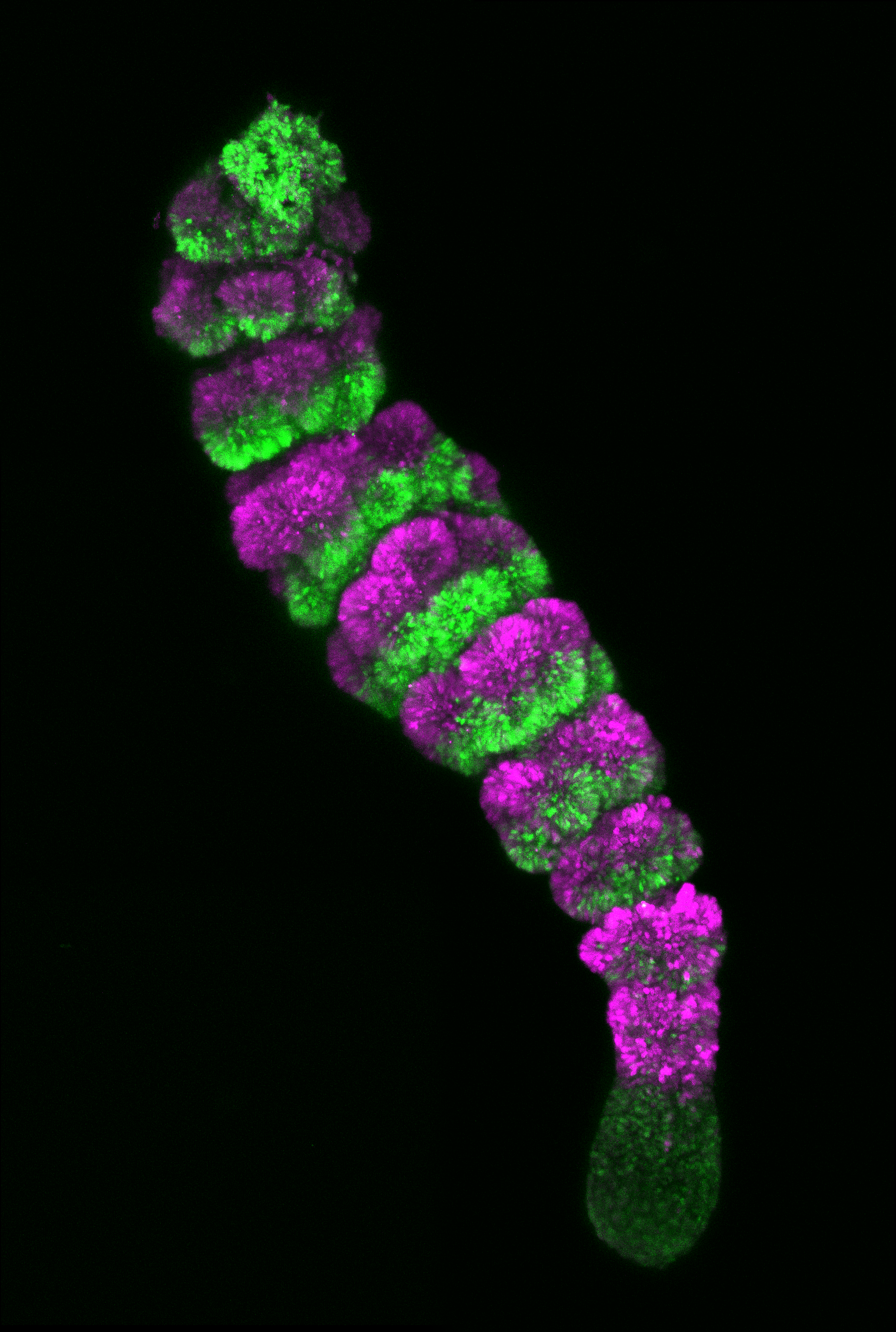
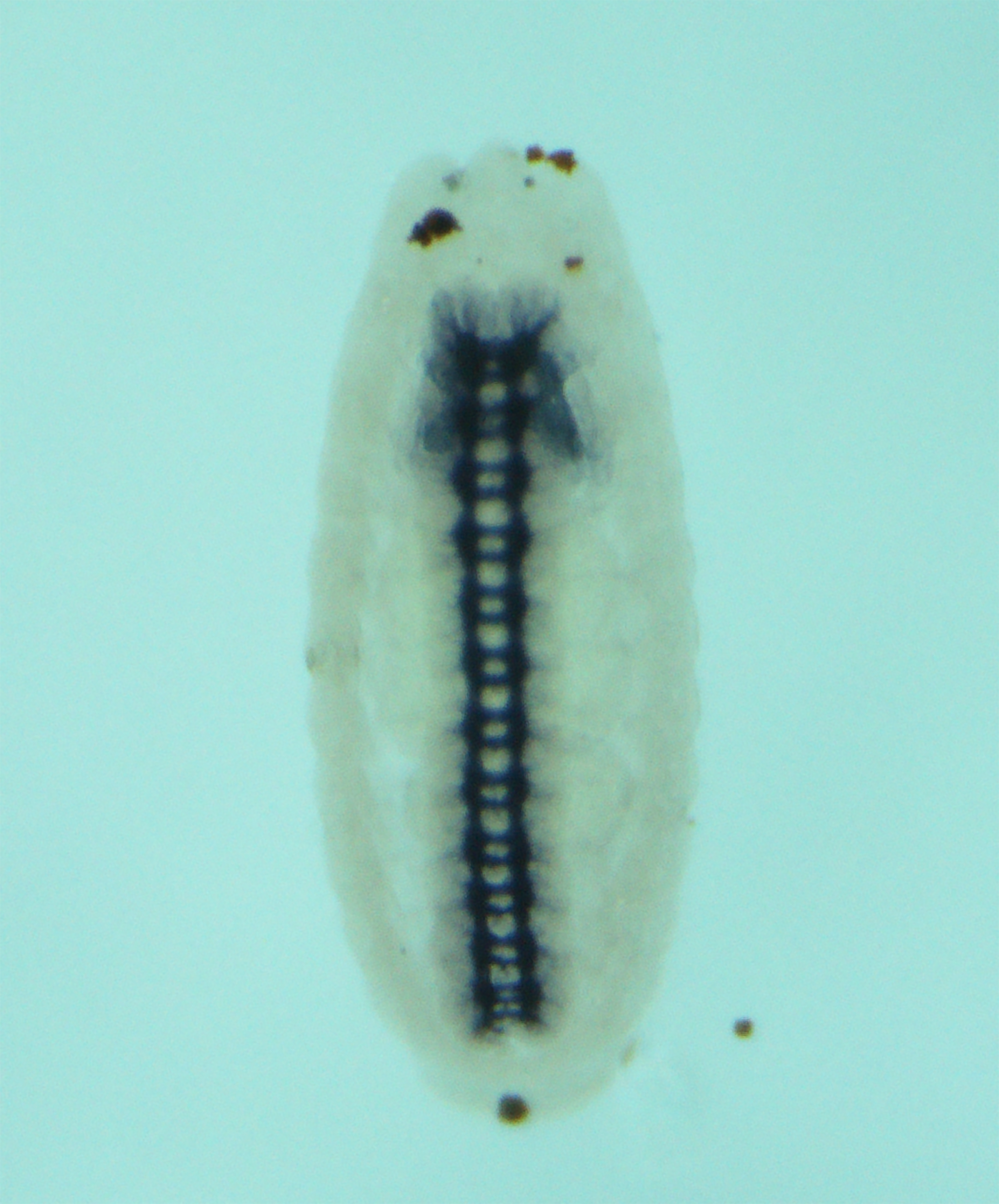
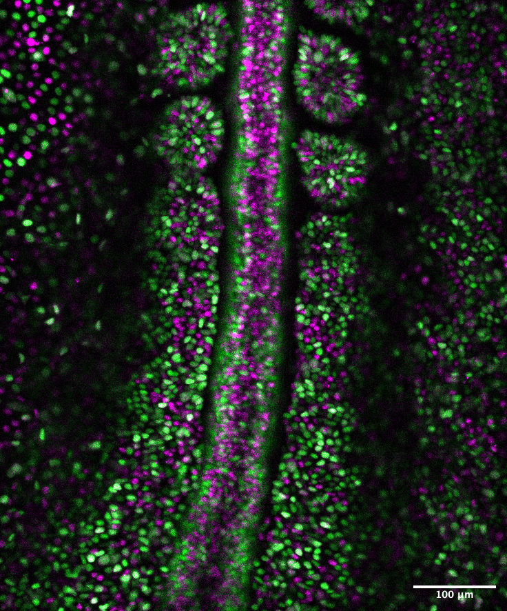
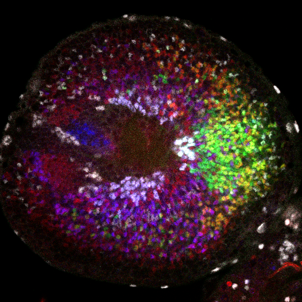
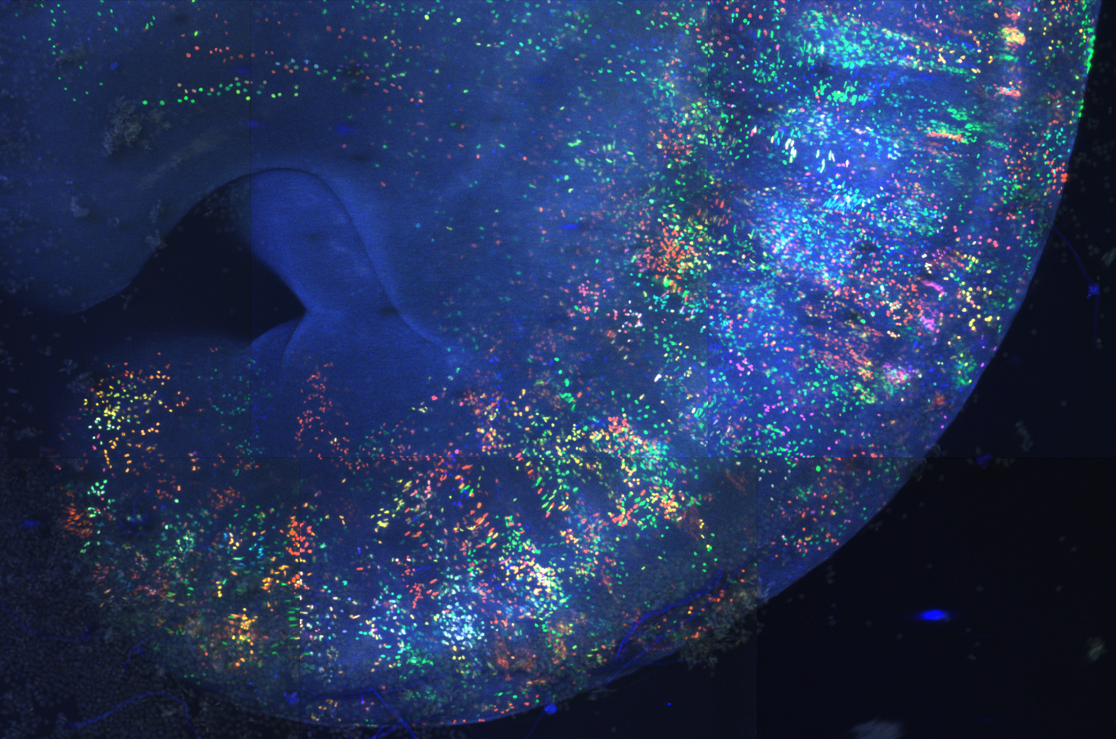
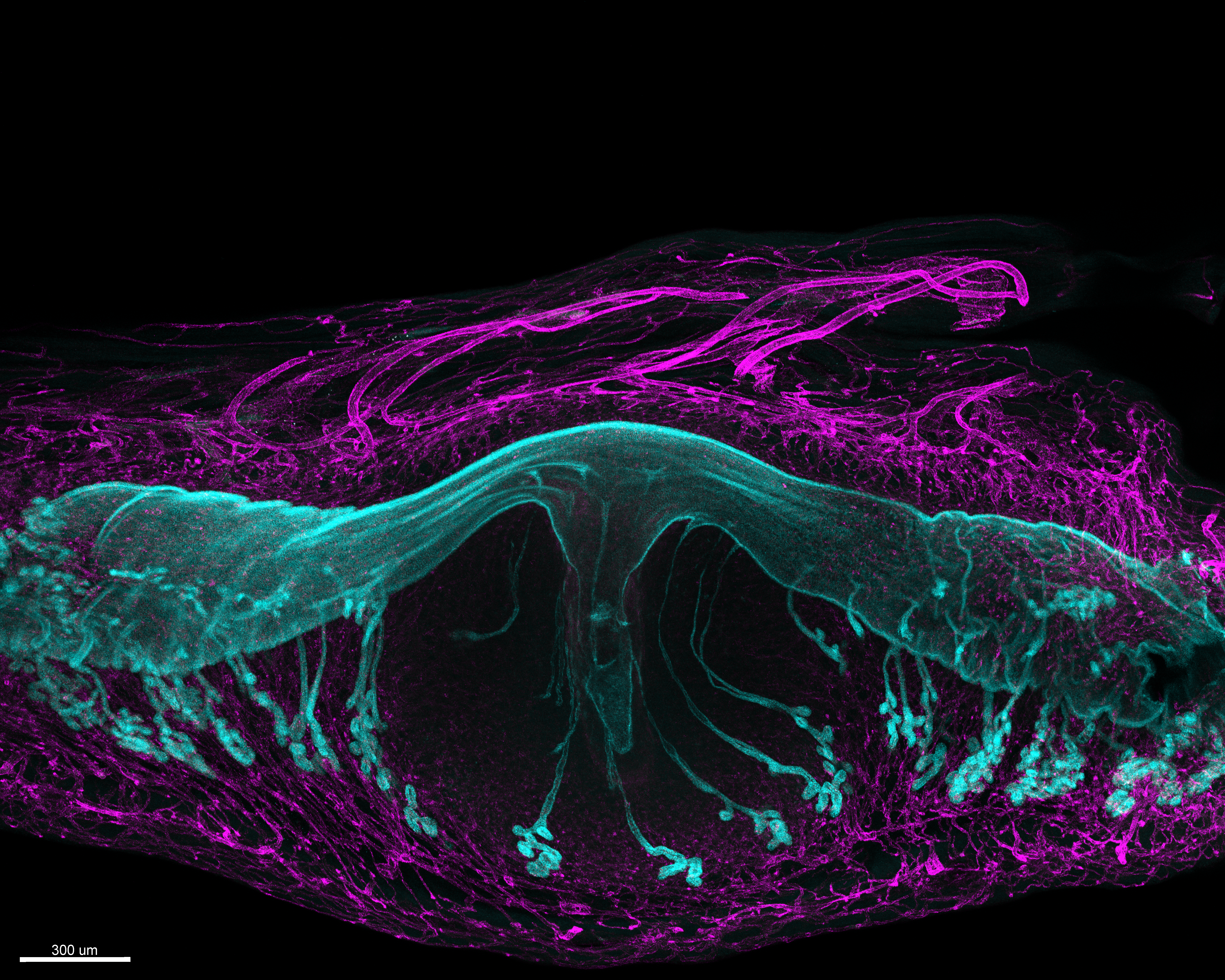
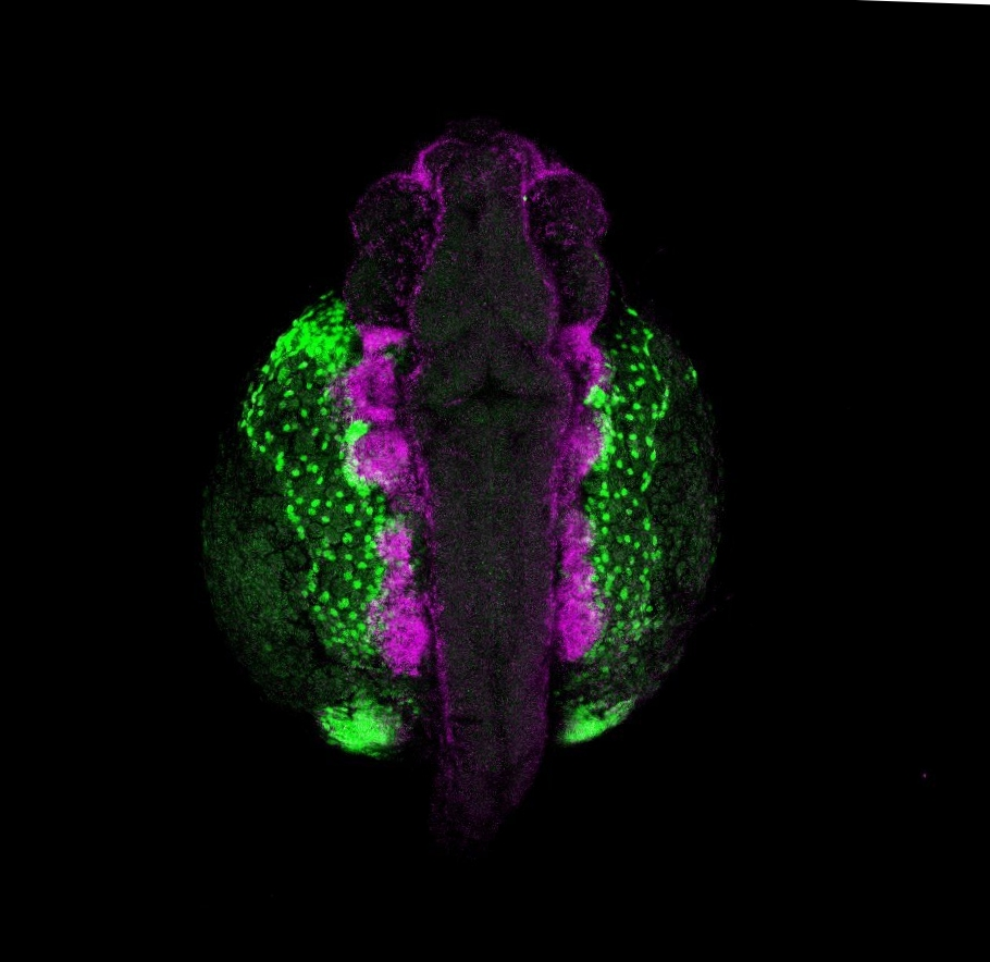
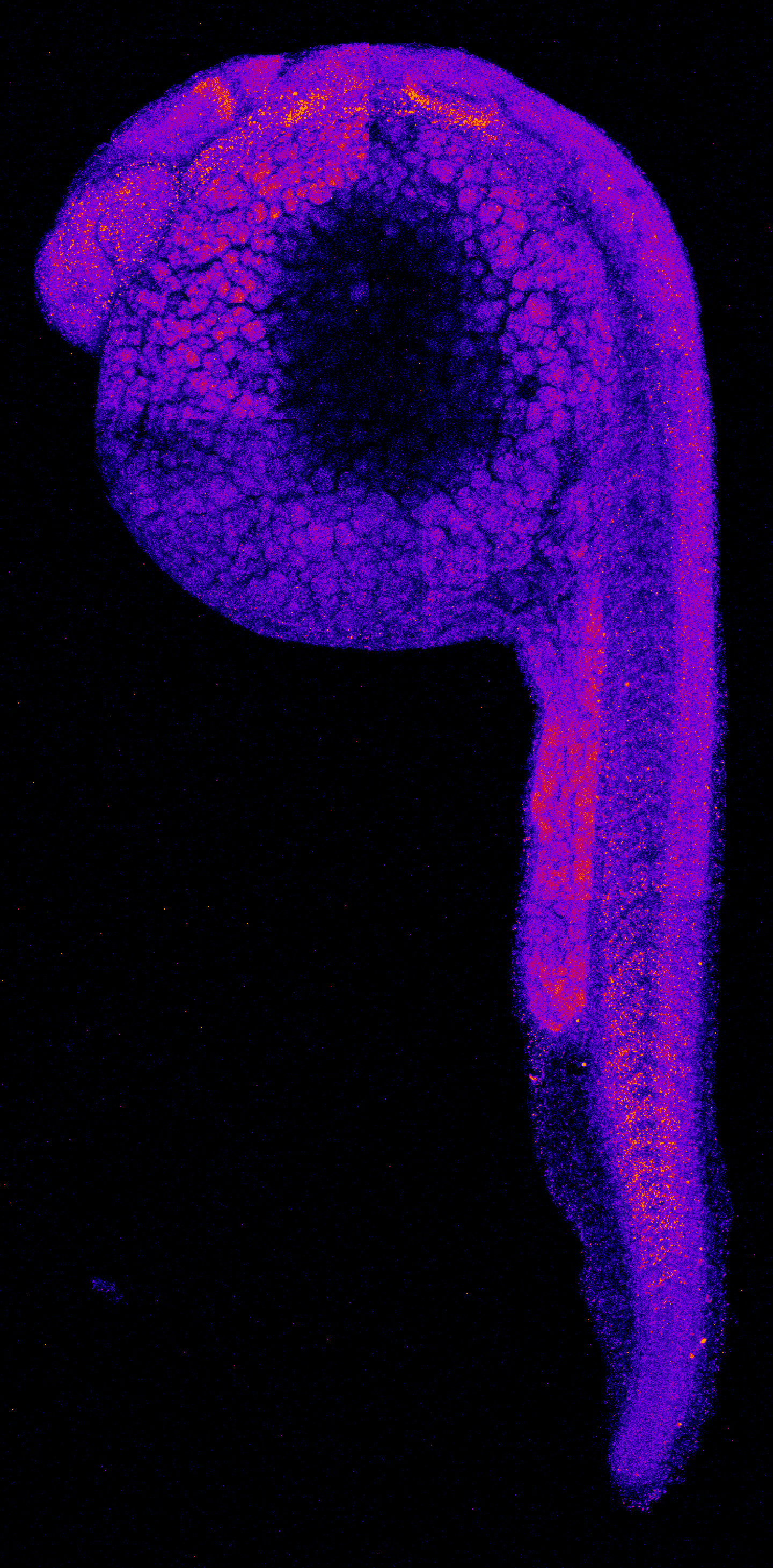
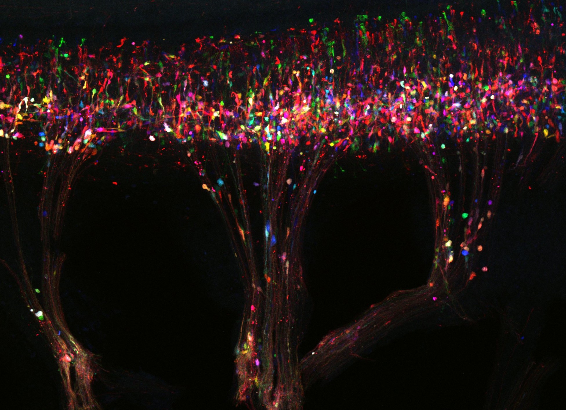
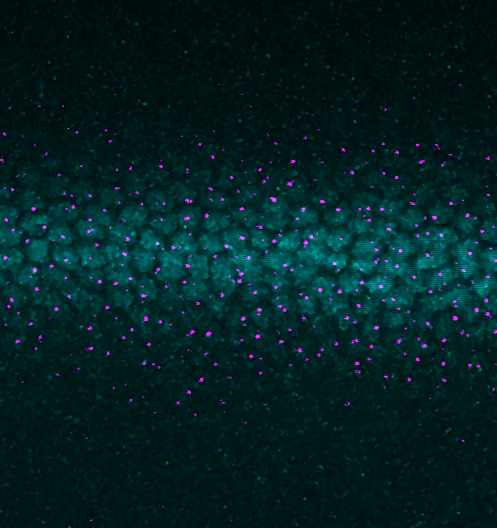
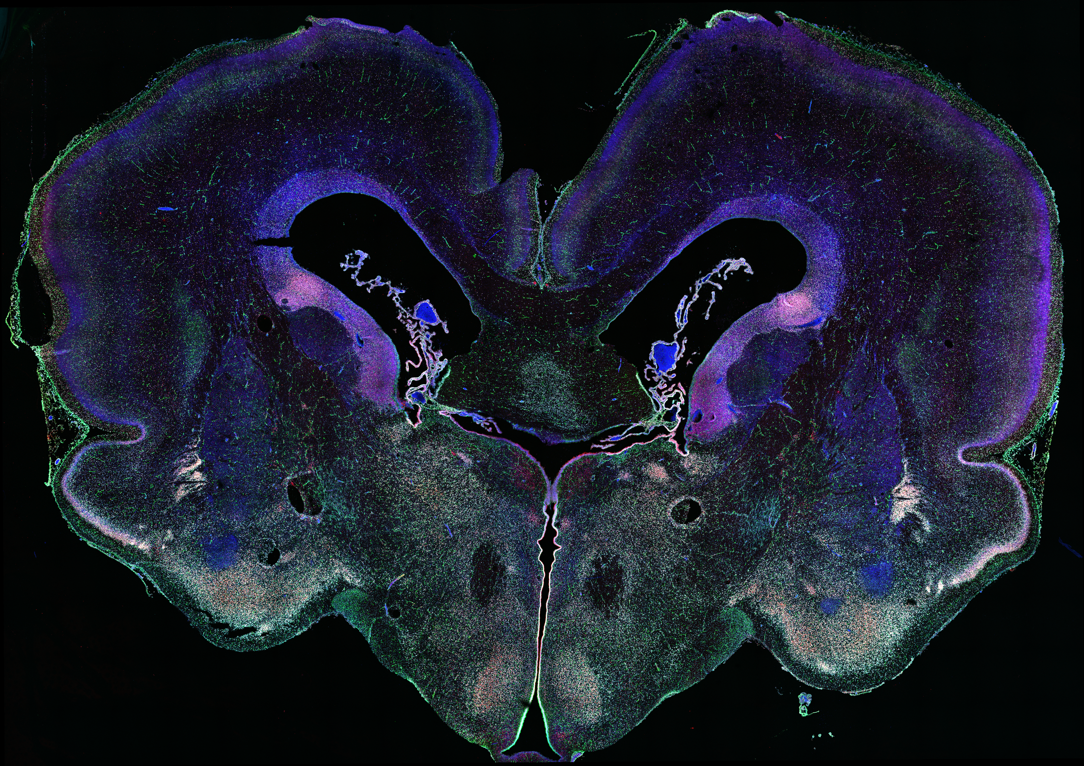
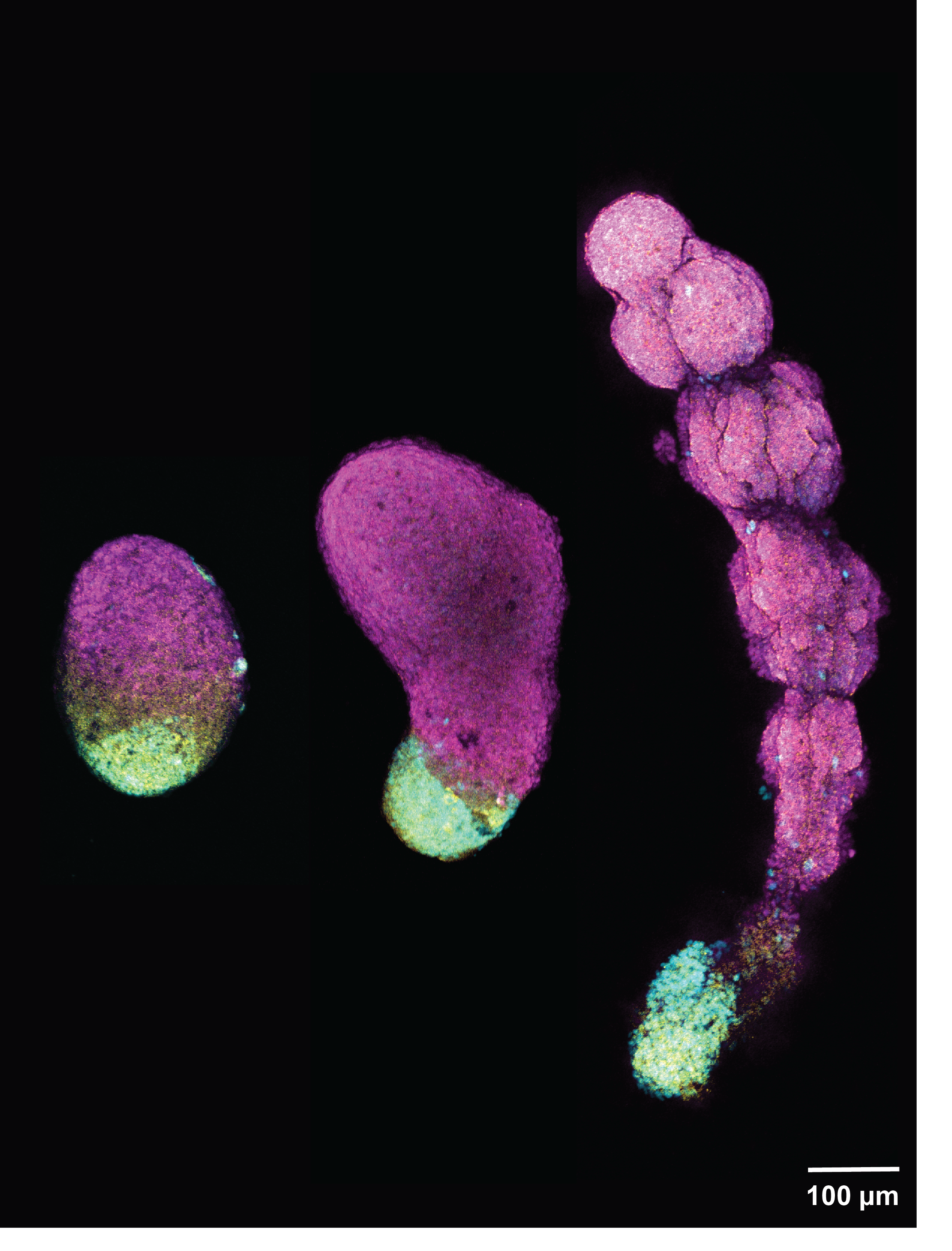
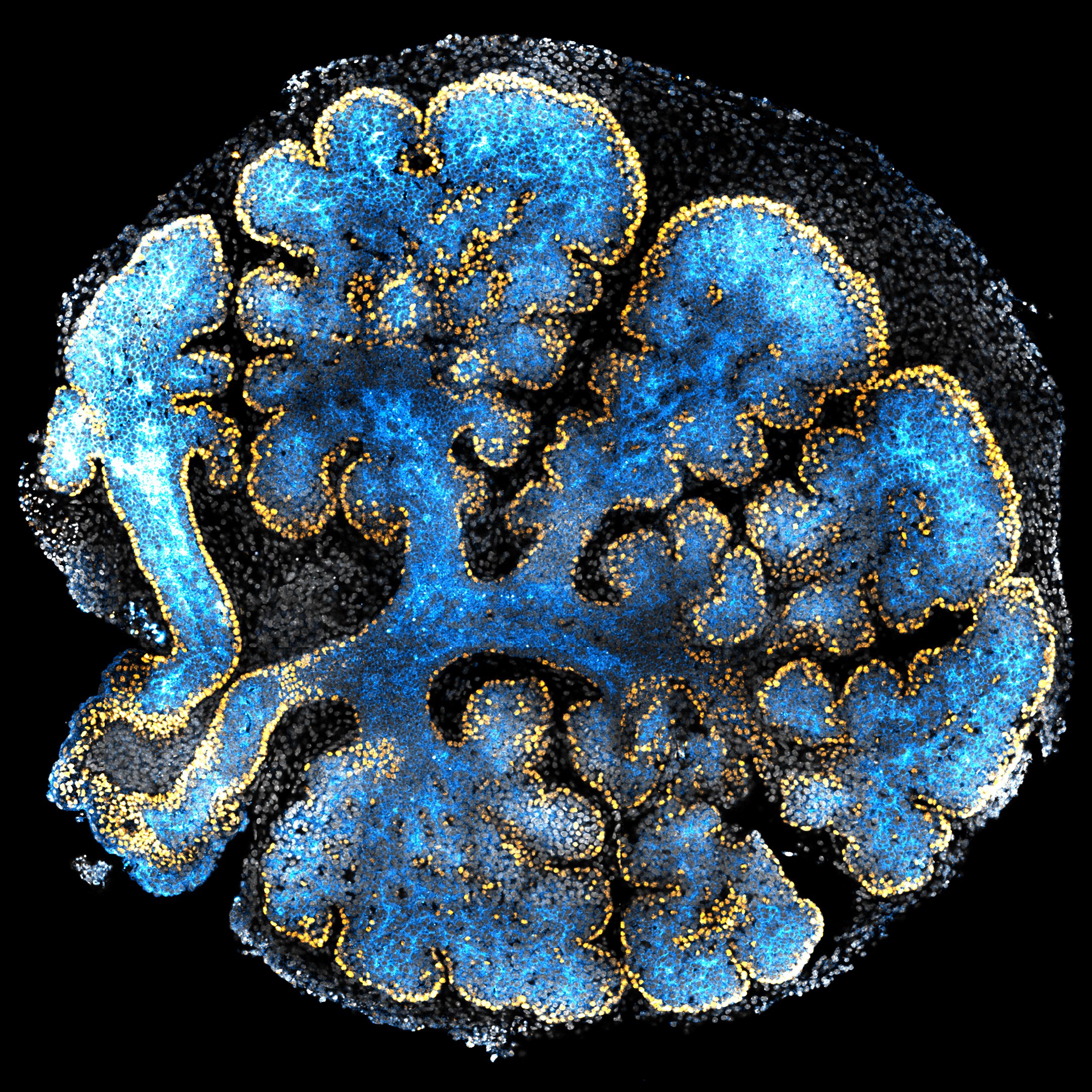
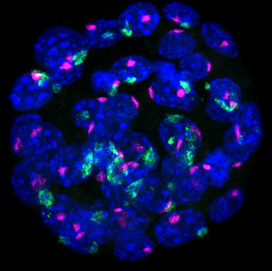
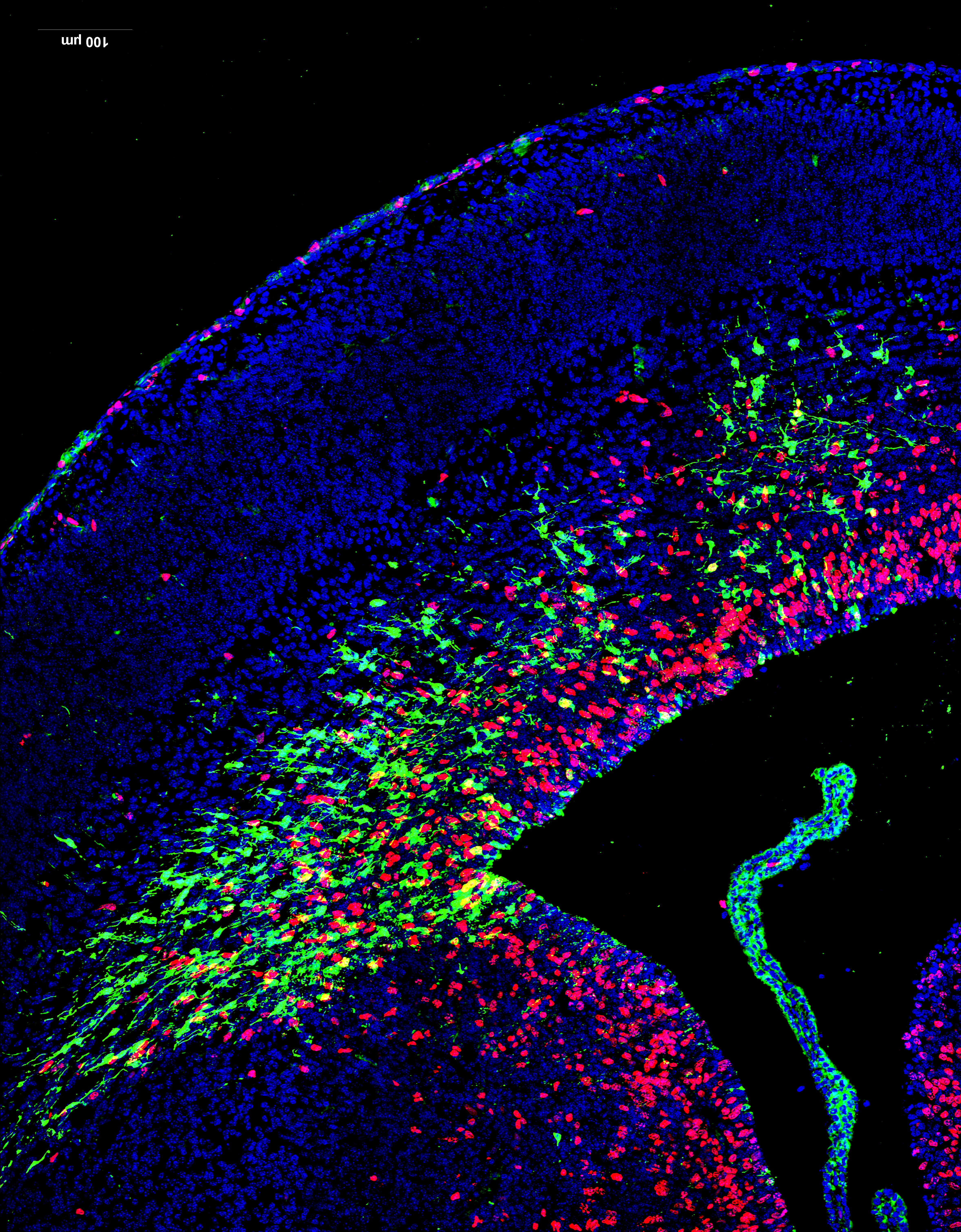
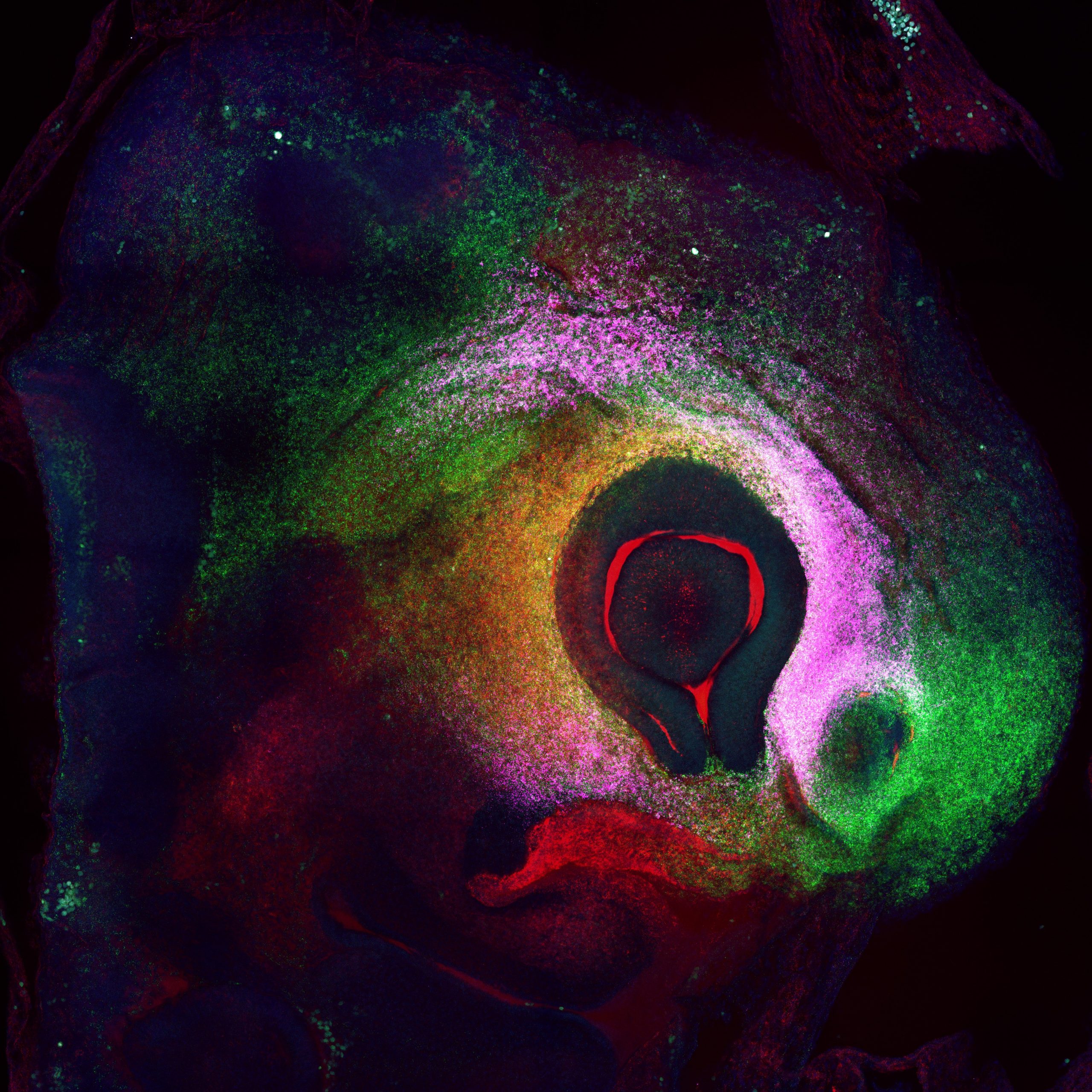
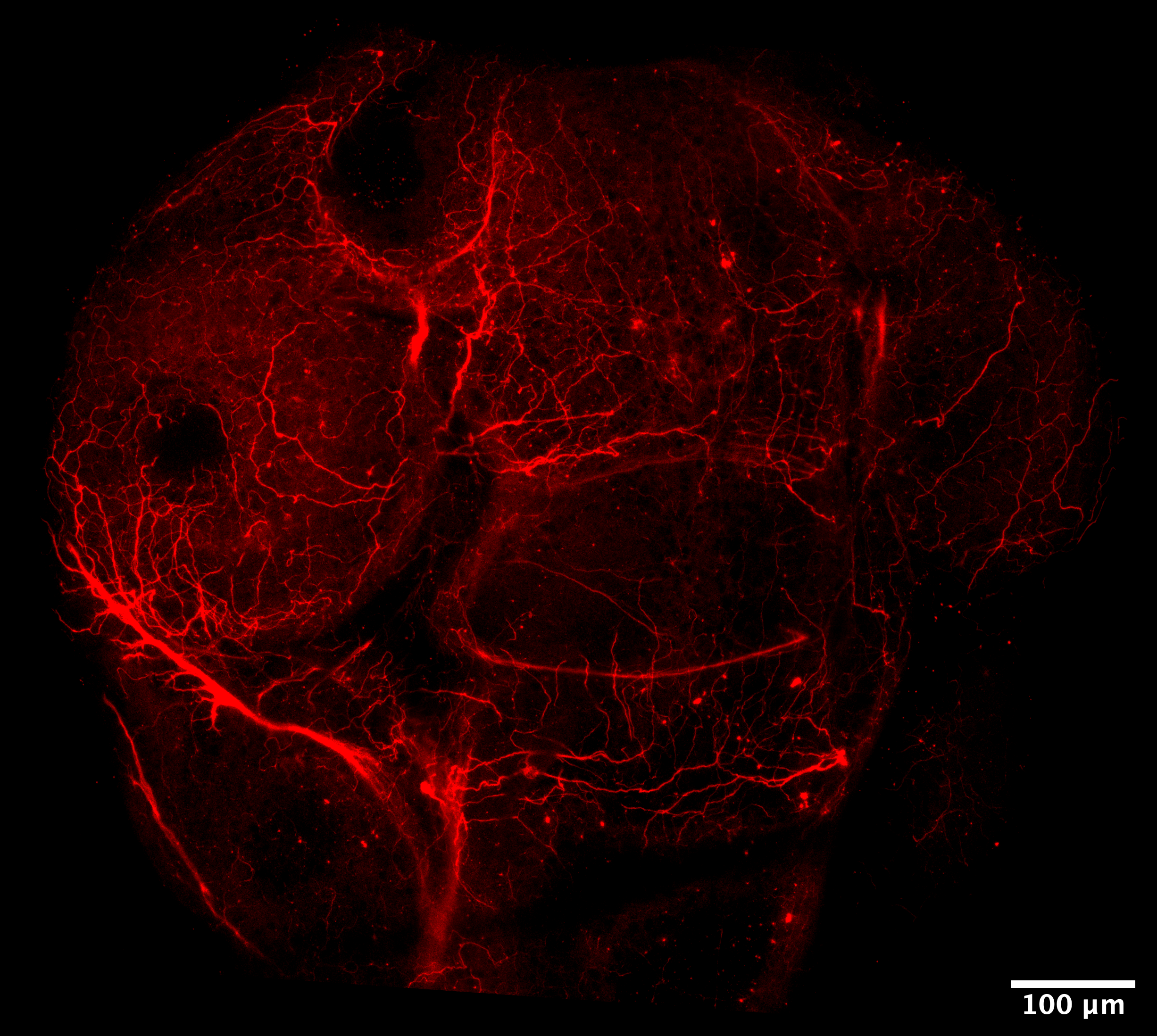
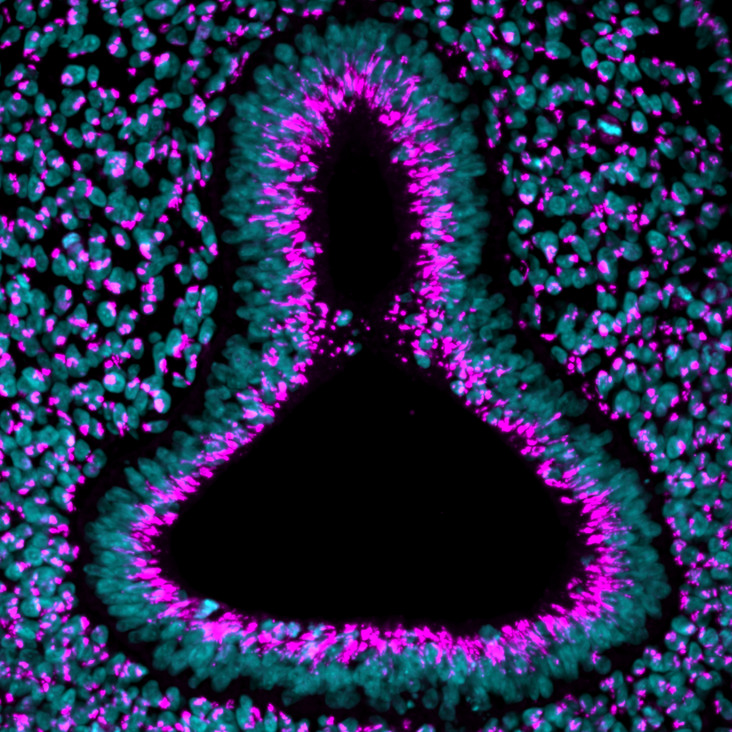
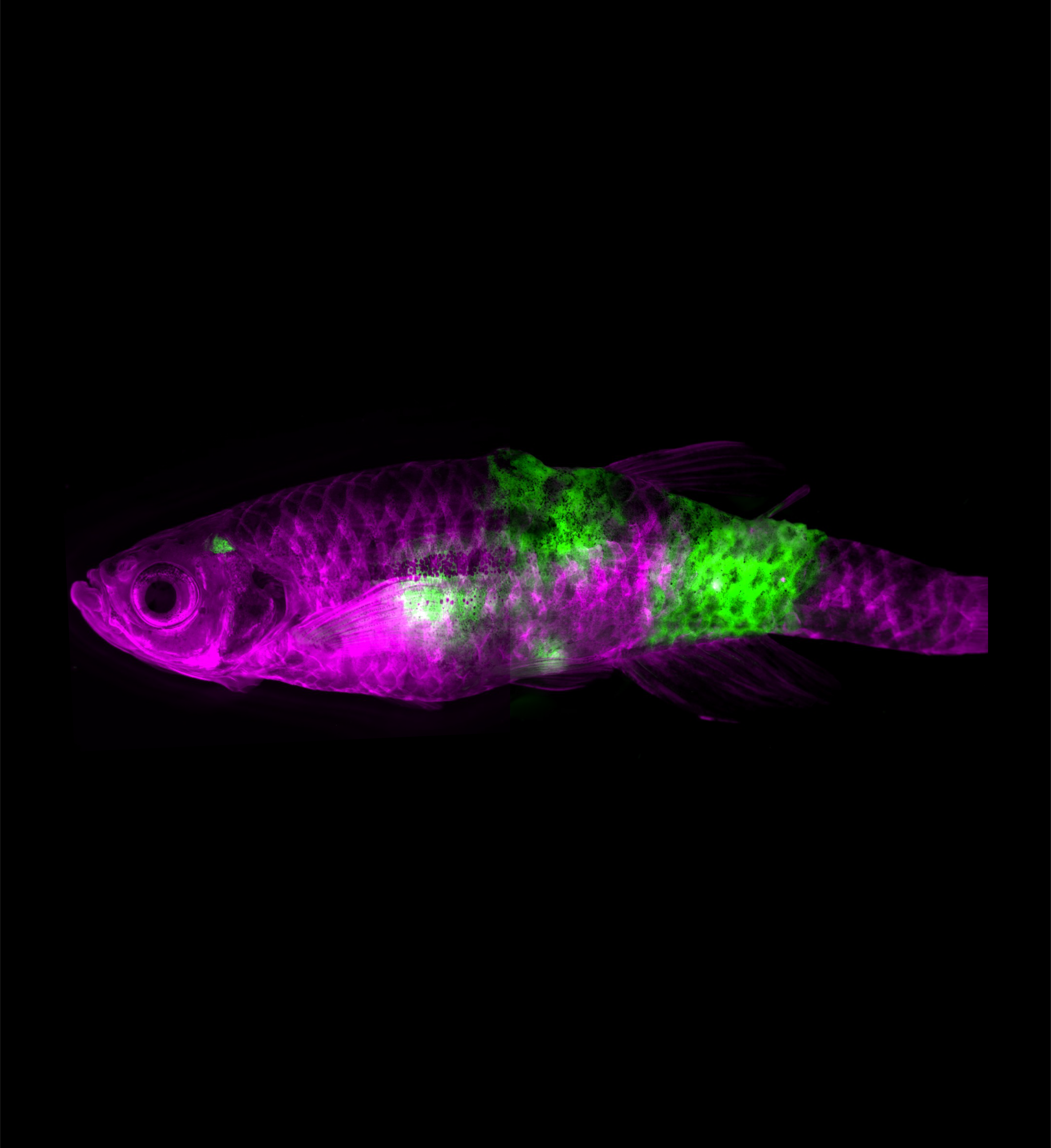
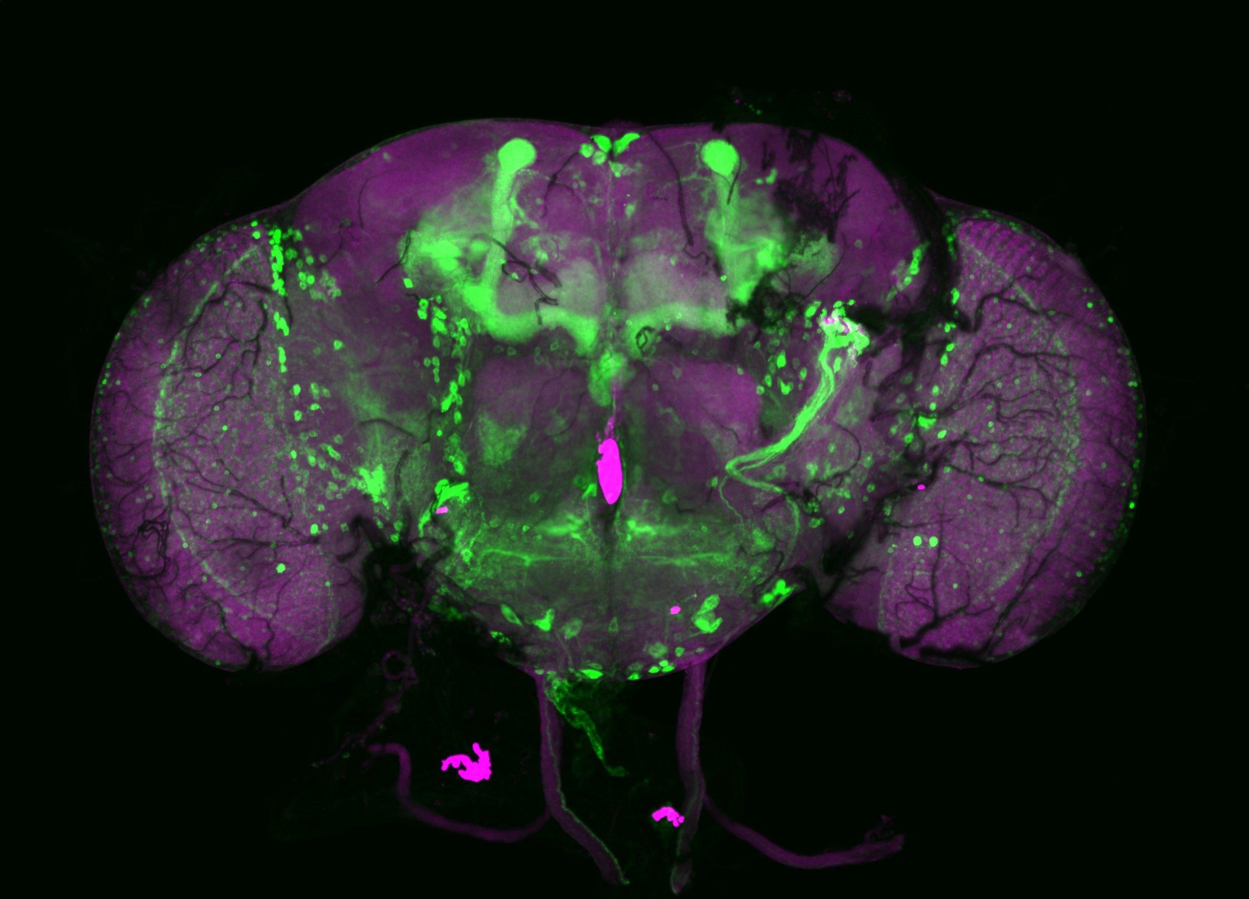
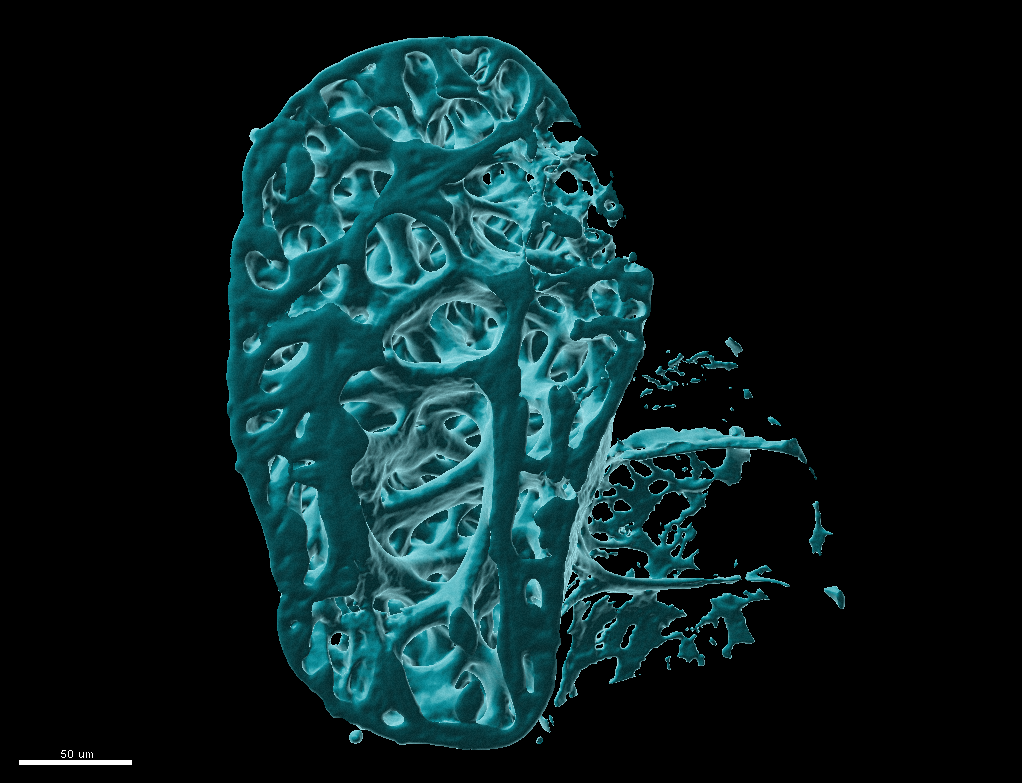
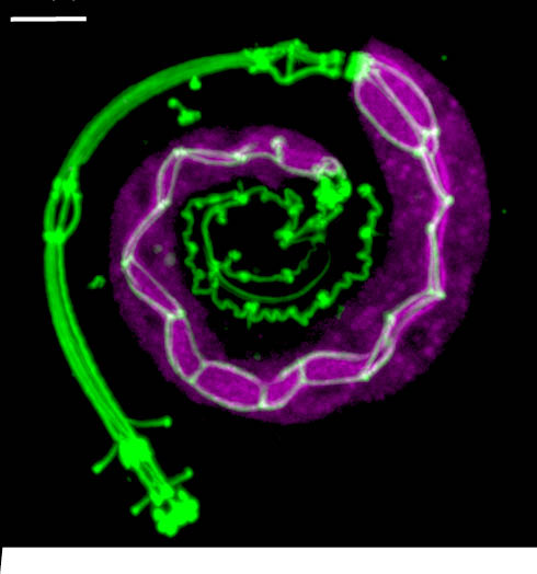
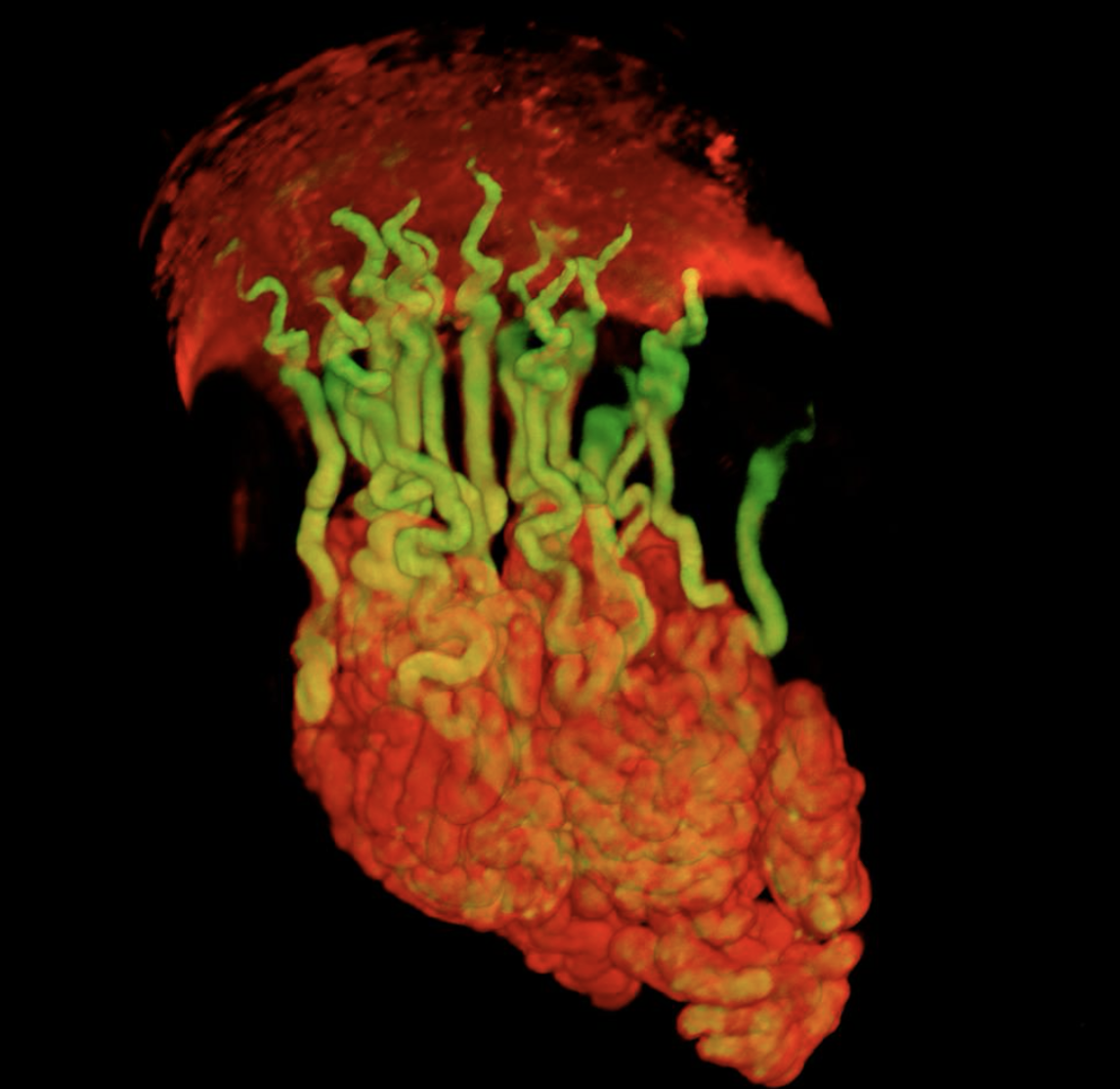
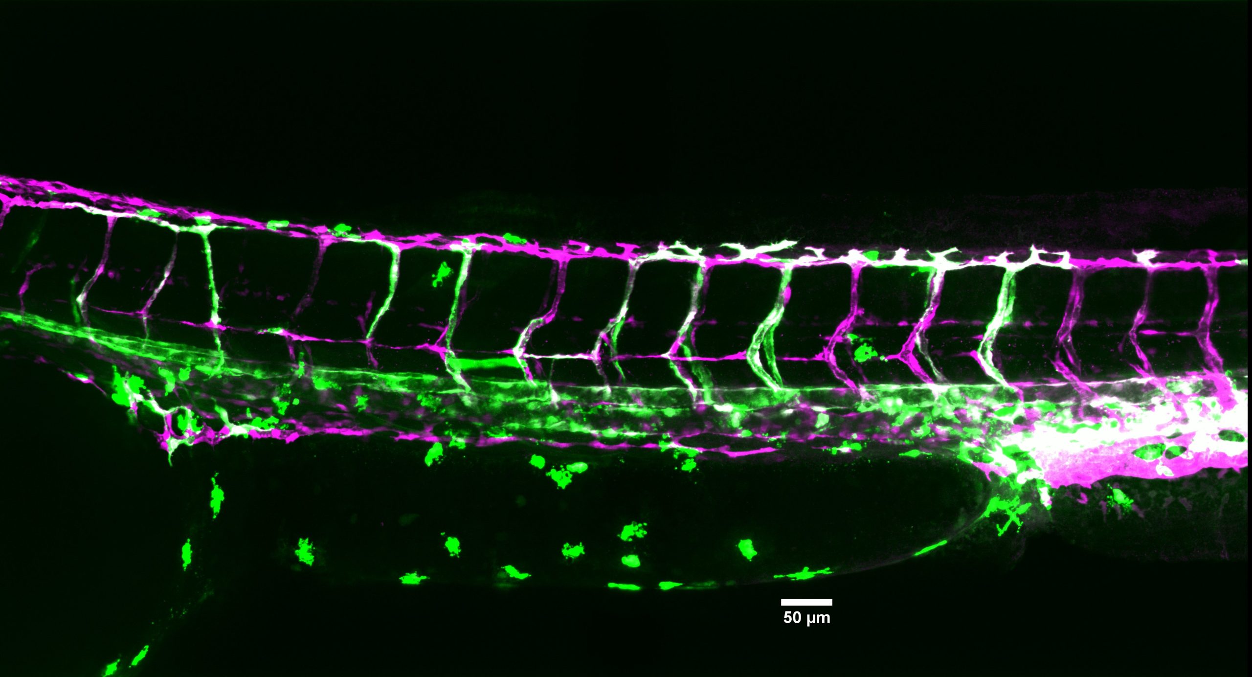
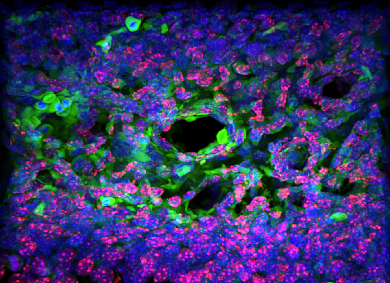
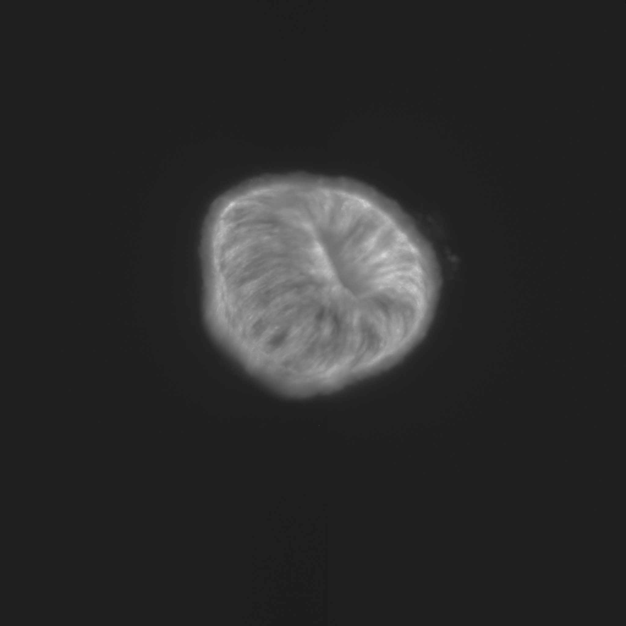
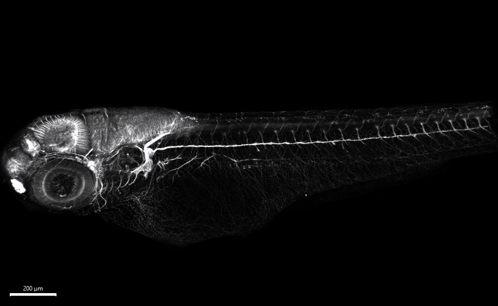
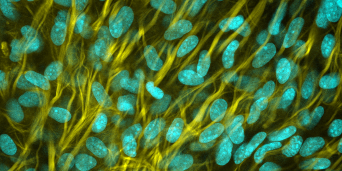
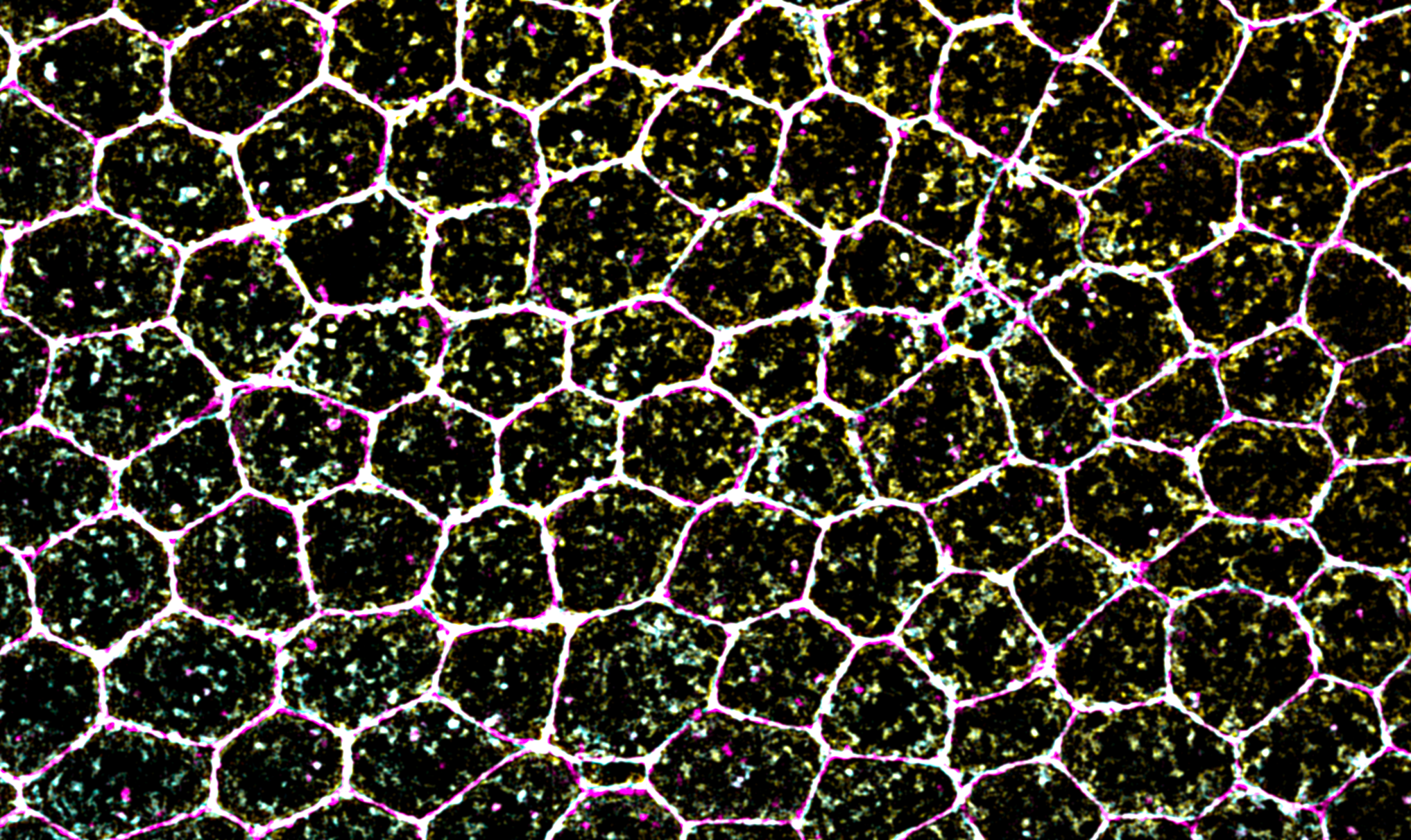
“Life as a drop in the ocean” This image is from an embryo of the marine acoel worm Hofstenia miamia, with cell membranes and nuclei highlighted. Early in development nuclei are shaped as rosettes but reasons or consequences for this remain mysterious. – Paul Bump“Human segmentoid” Organoids recapitulating human axial elongation and segmentation derived from induced pluripotent stem cells – Yuchuan Miao“Drosophila embryo Zipper” The Drosophila embryo central nervous system (CNS) stained with anti-body BP102 – Huangping An“Cycling through the cell cycle” Somite formation in chicken embryos. The pre-somitic mesoderm, the somites and the notochord are clearly visible, as the embryo grows towards the posterior, the anterior tissues mature. FUCCHI chicken line developed at the NARF and the Roslin Institute. Green marks cells in G2/M, Red marks cells in G1. – Ana R. Hernandez Rodriguez“Developing Neural Kaleidoscope in Drosophila melanogaster” Neuronal markers (TfAP-2 (red); Vsx1 (green); Ap (blue)) in the larval optic lobe of Drosophila melanogaster. Imaging was done with a Leica SP8 confocal microscope and processed with the LAS X and Fiji imaging software. – Yen-Chung Chen“Chicken Aurora” Tail of chicken embryo electroporated with Brainbow plasmids – Prikshit“Implantation Infrastructure: Uterine Glands and Vessels around the mouse embryo implantation chamber” Uterine Glands (Blue branched structures) and Blood Vessels (Pink) surround the elongating V-shape embryo implantation chamber (in the center) and are critical for supporting early pregnancy in the mouse. – Harini Raghu Kumar (Arora Lab)“A detour on the path to reproductive system morphogenesis in C. elegans” A young adult hermaphrodite C. elegans expressing markers for the somatic gonad (green) and germ cells (magenta) and carrying a mutation causing turning defects in gonad morphogenesis. – Noor Singh“Lateral neighbors” Developing zebrafish embryo showing the expression patterns of twist1 (magenta) and hand2 (green), dorsal view, anterior to the top. – Amanda Garfield“Fiery fish” A fire filter applied to a gene expressed in neurons and muscles along with DAPI to stain nuclei in a 24 hours old zebrafish embryo. – Zainab Afzal“Multichromatic Acacia” E6 chick embryo spinal cord electroporated with next generation Brainbow transgenes. This whole-mount acquisition allows tracing of neural progenitors to migrating neurons, following with axonal projections, showcasing lineage dynamics during development. – Thea Chrysostomou“Gradients” This image reveals the spatial coordination of BMP signaling and gene expression during early embryonic development. Dorsal view of a Drosophila melanogaster embryo at nuclear cycle 14, stained with anti-pSMAD antibody and smFISH for a BMP target gene. Captured using a Leica Stellaris confocal microscope and post-processed in Fiji. – Susanna Brantley“Symmetry on the Brain” This is a coronal section of a mid gestational embryonic piglet brain stained with DAPI in dark blue, phosphorlated s6 kinase in green, PROX1 in red, and NR2F2 in cyan to label different neuronal progenitor classes. Image aquired on a Leica Steallaris laser scanning confocal microscope then stitched and processed using ImageJ. – Emma Horton“Shaping it up like clockwork” Somite-forming organoids derived from human pluripotent stem cells allow the modelling of early vertebrate development and congenital disorders affecting the spine. Somites are transient block-like structures formed periodically, like clockwork, stacked on top of one another during early development, later developing into the vertebral column. The image shows a somitoid throughout its development from an oval spheroid to a stack of somites. Somite cells (magenta) develop on the anterior end, and progenitor populations (co-stained by cyan and yellow, which appear green) necessary for somite formation mark the posterior end. – Pranav S. Ramesh“Salivary cauliflower” Mouse embryonic salivary gland stained for Hes1 (blue) and p63 (yellow). Imaged using a 40x objective on a Zeiss LSM780 on the PICT Core facility at Institut Curie, Paris. – Robin Journot“Fishing for sex chromosomes” The X chromosomes (green) and Y chromosomes (magenta) painted by DNA FISH in a E3.5 mouse blastocyst; DNA stained with Hoechst (blue). – Aurélien Courtois“Radially migrating cortical neurons” After being born, the cerebral cortical projection neurons migrate from the proliferative zone of developing cortex to destined cortical layers. This extended depth of focus image shows these radially migrating cortical neurons at embryonic day 17 which are labelled with GFP expressing plasmid through in-utero electroporation. The staining in red shows EdU positive cells. This image has been taken using AXR Nikon microscope at CEAF Confocal facility, IIT Kanpur, India. – Nitin Agnihotri“Craniofacial mesenchyme”Craniofacial mesenchyme of HH16 chick embryo stained for ALX1 (magenta), ALX4 (green), PITX2 (red) and DAPI (blue). Image acquired with Nikon confocal microscope and processed with NIS element and FIJI. – Shirley Ee Shan Liau“Neural connections in the developing frog head” Stage 40 Xenopus laevis head labeled with an anti-neurofilament antibody, highlighting several neuronal connections. Antibody used: DHSB 3A10 (AB_531874). – Sudipta Mukherjee“Coming Together” This image captures the process during which the embryonic foregut forms an epithelial septum to give rise to the esophagus and trachea. Cells coming together in the septum remodel their Golgi apparatus (magenta), losing the tubular morphology and apical localization to the nucleus (cyan) characteristic of other epithelial cells, and resemble the surrounding mesenchymal cells. – Rui Yan“An Ewing Fish” Ewing sarcoma development through NCCs reprogramming. a massive green tumor driven by the expression of human oncofusion in neural crest cells. This fish also has an ectopic extra-dorsal fin, demonstrating an intriguing connection between normal development and tumorigenesis. – Elena Vasileva“Love on the Brain” Maximum intensity projection of an adult D. melanogaster brain from a fly carrying a GAL4 insertion crossed to a UAS-mCD8::GFP fluorescent reporter, expressing membrane-targeted GFP (in green) stained against phalloidin (in magenta) showing that the GAL4 insertion drives expression in the mushroom body of the fly brain. – Anastasia Repouliou“Web of Life” 20 days old zebrafish heart ventricle, membranes of cardiomyocytes are marked by fluorescent protein, imaged at 40x zeiss 980 and image rendered on Imaris. – Kirti Gupta“C. elegans digestive tract” – Lauren Cote– Catherine Pei-Ju Lu“The Developing Vasculature in Zebrafish” – Elithabeth Jones“The saga of bone genes and proteins” – Pragati Shekhar“Toroidal Hydra” – Daniel Pearce– Lucia RivasSomites and PSM eletroporated in chicken embryo (Gallus gallus). – Nisia MartinsFast green image of collagen in yellow and nuclei stained with DAPI in the suture mesenchyme of the mouse E15.5 skull. – Sasha Degtyareva Actin cytoskeleton in a developing Drosophila embryo – Nilay Taneja

 (No Ratings Yet)