Book review: Fast-forward: the fourth dimension in development
Posted by Development Book Reviews, on 23 November 2011
Development issue 24 features several book reviews. Over the next few weeks, these book reviews will also appear here on the Node. In this first one, Elaine Dzierzak and Catherine Robin compare developmental biology to Star Trek in their review of “Imaging in Developmental Biology: A Laboratory Manual” (Edited by James Sharp and Rachel O. Wong)
(Originally in Development.)
Book info:
Imaging in Developmental Biology: A Laboratory Manual Edited by James Sharp, Rachel O. Wong Series Editor, Rafael Yuste Cold Spring Harbor Laboratory Press (2011) 883 Pages ISBN 978-0-879699-40-6 (paperback), 978-0-879699-39-0 (hardback) $165 (paperback), $246 (hardback)
 Development is a bit like Star Trek, the long-running television series in which ‘space’ is the final frontier. For development, the final frontier is the fourth dimension, ‘time’. Time travel through the embryo, from the zygote to gastrulation, to organogenesis, and birth, has been a subject of fascination and science (fiction?) for centuries. This fascination is reflected in the many historical drawings of developing embryos and by advances in the field of embryology that came with the invention of the microscope. With the aid of microscopy, the field advanced from drawings of embryos to static images of fixed sections, which could be rendered, with some mental effort, into three-dimensional (3D) structures. However, comparisons of embryos at different formative stages could only hint at the patterns of dynamic cell growth and morphological change that occur during development, which recent molecular and genetic analyses have begun to uncover. Importantly, the current advances being made in innovative, real-time imaging technologies and in the computational processing of images have now fast-forwarded the field boldly into the dynamic fourth dimension. These advances are now summarized and explained in a newly published book on imaging, Imaging in Developmental Biology, edited by James Sharp and Rachel O. Wong, both experts in this field.
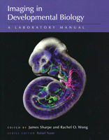
Imaging in Developmental Biology is an excellent resource from which both novices and experienced researchers can obtain current state-of-the-art embryo-imaging protocols for studying key developmental events, such as cell-fate determination, morphogen gradient formation, cell-cell interactions, cell migration and morphogenesis. The eye-catching cover immediately attracted passing lab members, encouraging them to browse the book, which they did with increasing interest. The first comment often expressed was: “I did not know that we could do so much!” Upon first perusal, this comprehensive book seems almost overwhelming with an impressive 57 chapters and seven appendices. But it does contain just about everything known about imaging embryos. This is not surprising as the volume is based, in part, on the popular and excellent Cold Spring Harbor imaging course. The editors have organized the book into four large sections, which contain chapters that are frequently and conveniently cross-referenced. A particularly helpful table is provided in Chapter 1 that guides the reader to specific protocols of interest in different animal models.
Section 1 (Chapters 1-7) provides a general entry into the imaging of common model organisms. This section’s emphasis on the advantages and disadvantages of each model organism allows researchers to evaluate quickly which animal model and procedures might be most useful for their specific investigations. Information is provided on embryo accessibility and size, tissue transparency, different cell-marking techniques, phototoxicity, mounting methods and compatibility with imaging resolution, and image analysis. Chapters are written by experts in the field, and each starts with a brief introduction into the mechanisms of development being addressed and with a description of the specific imaging method used, followed by one or more protocols. Concise troubleshooting, discussion, recipes, web resources and reference sections provided at the end of each chapter are of added benefit in ensuring the success of the approach.
Live imaging of cells is the focus of Section 2. Cell-labeling protocols in Chapters 8-13 provide an extensive coverage of dye injection and electroporation techniques for groups of, and for single, cells in chick, mouse, Xenopus and zebrafish embryos. The protocols provide important technical details about, for example, injection pipette production, electrode placement, voltage strength, pulse duration and intervals. Other labeling techniques covered include the difficult MARCM (mosaic analysis with repressible cell marker) in Drosophila and MADM (mosaic analysis with double marker) in mice. Commonly used direct transgenesis and knock-in strategies with fluorescent marker genes, as well as Cre-Lox recombination and fluorescence-photoactivation methods, are presented in the context of specific developmental questions concerning cell-lineage tracing. The relative merits of the different strategies are clearly described in Chapter 13. Chapters 14-23 cover live cell imaging of cell migration, mostly of the neuronal system. Particularly useful here are the descriptions of the use of the photoconvertible fluorescence marker Kaede to study neuron birth dating in zebrafish embryos, and the explanation of DNA injection methods, embryo embedding, image acquisition and software usage in the context of studies of zebrafish retina and lateral line development. As mouse developmental biologists, we found the zebrafish protocols and figures (in Chapters 16-18) easy to understand. Of special interest to us were Chapters 20 and 21, as the details of pre- and post-gastrulation mouse embryo dissections with excellent accompanying figures are an important resource for our students. These chapters contain detailed protocols for tagging cells with dyes, grafting cells into embryos and for electroporating DNA into groups of cells. The excellent figures and descriptions that detail how to culture embryos, hold them in place and manipulate them will be of great use for studying cell-fate determination and migration in early mouse development. Chapters 19 and 23 describe the imaging of embryo slice cultures in various species and tissues. As an important aspect of neural development, the imaging of circuit formation is the focus of Chapters 24-35, which are aimed at specialists studying neuron excitation, axon pathfinding or synapse formation. However, these approaches are also of broader relevance to research in other developmental systems.
Imaging multicellular tissues and organs as a whole dynamic entity is covered in Section 3. The first two chapters (Chapters 36 and 37) explain how to quantify morphogen gradients over time in Drosophila embryos. We found these chapters harder to understand and aimed more at the specialist reader. By contrast, Chapters 38-43 on cell movement describe more commonly used methods to acquire high-resolution dynamic movies of cells or subcellular proteins during morphogenesis in Xenopus, zebrafish, chick, quail and mouse embryos. Protocols provided in this section include how to generate Xenopus and zebrafish embryos with mosaic fluorescent reporter expression and how to prepare and mount tissue explants or whole embryos for long-term imaging, together with useful troubleshooting tips. This section also contains a well-illustrated protocol to perform high-resolution multiphoton time-lapse imaging of a developing chick embryo and protocols for 3D time-lapse imaging of epithelial morphogenesis and of the mouse metanephric kidney. Two relatively new imaging technologies are presented in Chapters 44-46 that allow live imaging of developing whole embryonic organs – optical coherence tomography (OCT, an optical version of ultrasound used to image high-speed events) and a non-invasive ultrasound technology used to image 3D organ geometry (which allows developmental events to be imaged over time).
After single cells and organs, Section 4 focuses on imaging the whole embryo in 3D and 4D. These chapters provide detailed protocols for the collection of spatial and temporal patterns of gene expression, for embryo phenotyping and for generating static or dynamic 3D atlases of model systems. Of particular interest is Chapter 48, which provides a didactic description of the intellectual and technical efforts of the Berkeley Drosophila transcription network project, which has produced a computationally analyzable, gene expression and morphological 3D atlas of a blastoderm embryo. The use of hardware and software is also well explained and illustrated in detail, including chapters on optical projection tomography (OPT), X-ray micro-computed tomography (μCT), episcopic fluorescence image capturing (EFIC), high-resolution episcopic microscopy (HREM) and macroscopic magnetic resonance imaging (μMRI), with detailed information again provided for troubleshooting as well as additional web resources. Descriptions of the advantages, disadvantages, limitations and future prospects of each technique will definitely assist readers in choosing the appropriate method for their own application. The fourth dimension, time, is finally reached in the last chapters (56 and 57). Protocols are provided here for high-volume 3D time-lapse imaging of live adult Caenorhabditis elegans and of zebrafish and Drosophila embryos. Digital representations of these embryos permit cell tracking in time, revealing their origin and fate, which is of particular interest to developmental biologists.
Altogether, Imaging in Developmental Biology is a book for those Development readers who are curious to know more about new technical developments and possibilities within the exciting field of imaging. It is a valuable guide and a very helpful laboratory manual for students, although serious training might be required to perform the more complex experiments. Seven appendices at the end of the book are particularly helpful for new people entering the field and include a must-have list of fluorescent filters and excitation/emission spectra, lens cleaning tips, a list of cautions and potential disasters, and an all-important glossary of terms. The book is pleasant to read, with its clever use of illustrations, photos and online protocol videos. Thus, we highly recommend this book and hope that multidisciplinary collaborative expertise in biology, imaging, image analysis, computer science, visualization and database construction will continue to fast-forward 4D imaging techniques and, consequently, our knowledge of development.


 (6 votes)