Featured image with Özge Özgüç: the Node–FocalPlane image competition
Posted by the Node, on 6 May 2025
To accompany the Biologists @ 100 conference, we’ve partnered with FocalPlane to bring to you an image competition. We shortlisted 15 images and asked you to vote for your favourite online and in person at the conference. On the final day of the conference, we announced the top3 images of the competition.
In this ‘Featured image’ post, we find out more about the story behind Özge Özgüç’s image, which was the winner of the competition.
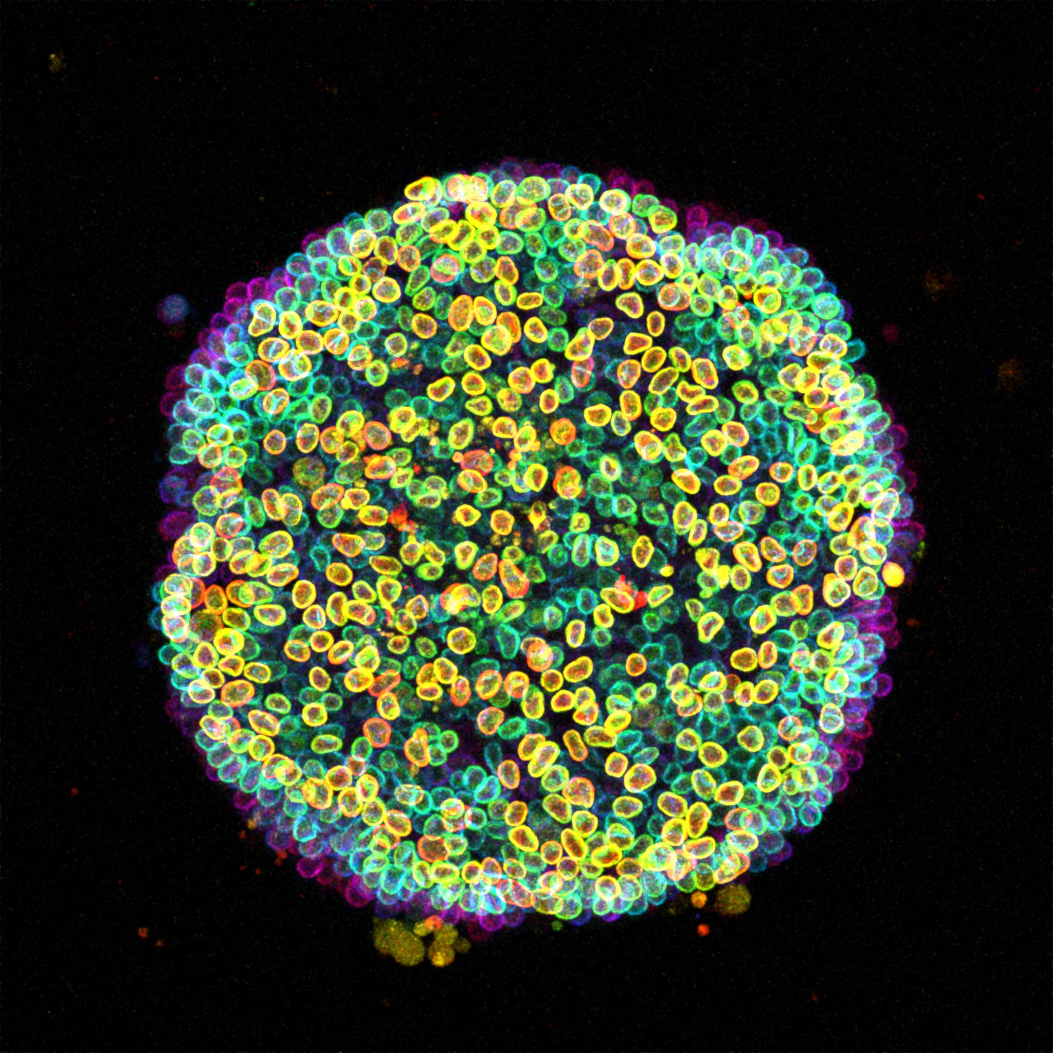
A ‘Cell-estial Bloom’ of human induced pluripotent stem cells (hiPSCs) flourishes on a micropatterned island. This image presents a colony of live hiPSCs, with fluorescently labeled Lamin B delineating the nuclear lamina within each cell. Acquired with a Zeiss LSM 880 Airyscan microscope, this maximum intensity projection is enhanced with depth-coded coloring to reveal the captivating three-dimensional landscape.
What is your background?
I completed my undergraduate education in the Department of Molecular Biology and Genetics at Middle East Technical University (METU) in Ankara, Turkey. During this time, I participated in research across different fields of biology through various internships to discover what truly excited me. It was during one of these internships that I realized I was most interested in developmental biology. To follow this interest, I pursued a master’s in Developmental Biology at Sorbonne University in Paris/France, followed by a PhD in Cellular and Developmental Biology at Institut Curie in Paris/France in the lab of Jean-Léon Maître. During my PhD, I focused on the physical forces that shape the preimplantation mouse embryo, particularly how actomyosin contractility prepare itself for morphogenesis by transitioning from an egg to an embryo state before it becomes the driving machinery behind the morphogenetic events of preimplantation development. Currently, I’m a postdoctoral researcher at the Institute for Bioengineering of Catalonia (IBEC) in Barcelona/Spain in the lab of Xavier Trepat. My work has grown increasingly interdisciplinary, bringing together developmental biology, biophysics, and bioengineering, to explore how mechanical forces influence early developmental processes across different model systems.
What are you currently working on?
Currently, I’m working on building experimental models that allow us to study the mechanical aspects of early human development. Human post-implantation stages are notoriously difficult to access and study in vivo, so we’re developing in vitro systems that recreate aspects of this development in a controlled and mechanically accessible way. With these tools, I aim to understand how physical forces, like pressure and tissue tension, influence key cell fate decisions and morphogenetic events, such as the symmetry breaking and start of gastrulation.
Can you tell us more about the story behind the image that you submitted to the image competition?
This image comes from a live-imaging session of micropatterned human induced pluripotent stem cells (hiPSCs), with fluorescent Lamin B marking the nuclear envelope. I was curious about how the cells were packing their nuclei into such a confined space, so I applied color code for the depth. Seeing nuclei at different height with a different color revealed the layered organization which was both informative and eye-catching. I first used the image as a cover slide for my lab meeting and got very nice comments about it, so I decided to submit it to the competition. The name “Cell-estial Bloom” actually came up while chatting with colleagues because we couldn’t decide whether it looked more like a flower or a galaxy.
What is your favourite technique?
I really enjoy live imaging. Something is very captivating about watching cells move and change shape in front of your own eyes and I find it incredibly satisfying to capture dynamic processes as they unfold. But I also love techniques that let you physically interact with cells and tissues. For example, during my PhD, I used various methods to change the cell size and shape, like aspirating them into micropipettes, fragmenting, fusing, or placing them into molds. These kinds of manipulations gave me a very hands-on understanding of how cells respond to mechanical cues. So, overall, I think I’m mostly excited by techniques that combine observation with gentle intervention, where you’re not just watching biology happen, but actively nudging it to reveal how it works.
What excites you the most in the field of developmental and stem cell biology?
What excites me the most is how the development of new tools and techniques open doors to explore developmental processes that once seemed out of reach. I love how these innovations often bring together ideas from completely different fields and invite you to look at the developmental processes from a fresh angle.
 (1 votes)
Get involved
Create an account or log in to post your story on the Node.
Sign up for emails
Subscribe to our mailing lists.