Vote for a Development cover – Woods Hole 2012 class round 1
Posted by the Node, on 30 January 2013
Each year, students of the Woods Hole Embryology course produce some amazing images. Last year, readers of the Node selected four images from the 2011 course to appear on the cover of Development.
Now it’s time to do the same with the images from the 2012 course. Here’s the first batch of four images. Please vote in the poll below the images for the one you would like to see on the cover of Development. (Click any of the images to see a bigger version.) Poll closes on February 19, noon GMT.
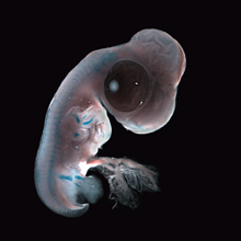
 1. Chick ectopic limb. An FGF-4-soaked bead was implanted at stage 14. The embryo was fixed four days later, and stained with alcian blue to reveal the developing cartilage of the skeleton. An ectopic limb can be seen developing next to the normal forelimb, and the bead is still present in the body wall. This image was taken by Elsie Place (MRC National Institute of Medical Research).
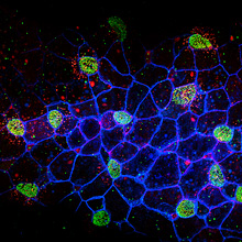
 2. Two day old Xenopus embryo epidermis, highlighting multiciliated cells. The embryo had been injected with mRNAs encoding membrane blue fluorescent protein, Centrin GFP, and Clamp RFP at the 4-cell stage and imaged as a live mount by confocal microscopy. This image was taken by Andrew Mathewson (Fred Hutchinson Cancer Research Center).
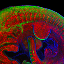
 3. Confocal image (extended focus Z stack) of an E10.5 day mouse embryo (lateral view; thorax) immunostained with antibodies against PECAM (endothelial factor present in the vasculature; red), beta-III-tubulin (neurons; green) and DAPI (cell nuclei; blue). This image was taken by Joyce Pieretti (University of Chicago), Manuela Truebano (Plymouth University), Saori Tani (Kobe University) and Daniela Di Bella (Fundacion Instituto Leloir)
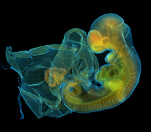
 4. Mouse embryo, day E9.5. Widefield fluorescence image showing immunostaining with anti-Tuj1 (orange) and anti-glucagon (green), counterstained with DAPI (cyan). This image was taken by Eduardo Zattara (University of Maryland, College Park).


 (7 votes)
Awesome images, kudos to the students!!
Indeed, lovely. Thanks for putting them all up for consideration!
Anyone have any idea why the apical ectodermal ridge stains with beta-III-tubulin? (Innocent question)