In Development this week (Vol. 142, Issue 2)
Posted by Seema Grewal, on 6 January 2015
Here are the highlights from the new issue of Development:
The ‘second brain’: taking gut development up a Notch
 The vertebrate gastro-intestinal (GI) tract consists of a regionalized epithelial tube surrounded by mesenchyme that later differentiates into smooth muscle. During the early stages of stomach patterning in chick embryos, the primitive GI track is colonized by vagal enteric neural crest cells (vENCCs), which will give rise to the enteric nervous system (ENS). The important role of the ENS in controlling GI function is well understood, but its contribution to the development of the GI tract has never been addressed. On p. 331, Sandrine Faure, Pascal de Santa Barbara and co-workers demonstrate that vENCC ablation impairs mesenchyme proliferation and differentiation. Moreover, reducing the number of vENCCs alters the molecular identity of both the mesenchyme and the epithelium, such that they express intestinal markers. Mechanistically, the authors show that these defects in stomach patterning and differentiation result from the ectopic activation of Notch and BMP4 signalling; the downregulation of both these pathways is necessary for proper stomach development. Altogether, this work reveals that vENCCs control stomach patterning and differentiation through the inhibition of Notch, shedding light onto the mechanisms that govern the contribution of the ENS to GI tract development.
Chickadee: building a nest for the germline
 The apical region of the adult Drosophila testis harbours a stem cell niche that contains germ stem cells, which differentiate into spermatocytes, and somatic cells, which provide nutrients and regulate the proliferation and differentiation of the germline. During spermatogenesis, somatic cells encapsulate the germline cells, isolating them from the environment by providing a permeability barrier. Disruption of either encapsulation or permeability barrier function has catastrophic effects on spermatogenesis, resulting in sterility. Here, Guy Tanentzapf and co-workers investigate the genetic determinants of soma-germline interactions, specifically during germline encapsulation (p. 268). Using a novel permeability assay, they show that encapsulation and the creation of a permeability barrier are actually two separate processes. Furthermore, disrupting the function of chickadee, the Drosophila ortholog of Profilin, causes altered encapsulation and consequent failure of the permeability barrier formation. Lastly, the authors demonstrate that the permeability barrier, which needs functional junctional proteins, is required to restrict the range of niche-derived BMP signalling. In summary, this work identifies Chic as a key regulator of the two distinct phases of soma-germline interactions during early spermatogenesis.
A key role for Wnt-mediated laminin synthesis in fin morphogenesis
 Limb and fin morphogenesis start with the formation of the apical ectodermal ridge (AER), an epithelial signalling centre that coordinates appendage development. Wnt signalling is required for AER induction and several extracellular matrix (ECM) components are necessary for proper limb formation. Mahendra Sonawane and colleagues (p. 320) set out to explore the mechanisms regulating ECM synthesis and the role of Wnt signalling during appendage development – using the zebrafish median fin as a model. They observe that cell morphology in the distal part of the AER is distinct from the rest of the appendage epithelium, and that these differences in morphology correlate with a gradient of Wnt. Mechanistically, canonical Wnt signalling modulates cell shape with spatiotemporal precision by regulating the expression of the ECM component laminin α5, which signals via integrin α3 to influence cell morphology. Finally, the authors show that those mechanisms are conserved in the pectoral fin. This study uncovers a novel mechanism in which canonical Wnt signalling controls laminin synthesis to regulate epithelial cell shapes and tissue morphology during vertebrate appendage development.
Regulating progenitor pools in the lung
 The secretory and multiciliated cells of the adult lung are constantly replenished by multipotent epithelial progenitors: the basal cells. Basal cells give rise to parabasal intermediate progenitors, which then terminally differentiate into ciliated or secretory cells. However, the specific molecular mechanisms governing the production of parabasal cells in the lung remain mysterious. Using genetic and pharmacological approaches in air-liquid interface cultures of adult airway progenitors, Wellington Cardoso and colleagues (p. 258) find that selective activation of Notch 3 identifies parabasal cells and controls the balance between basal and parabasal progenitor cells in airways. The authors show that Jagged 1 and 2 in basal cells are crucial for activation of Notch 3 signalling and for the generation of the pool of parabasal cells. Notably, individuals with chronic obstructive pulmonary disease were found to exhibit Notch 3 hypo-activation and an expanded basal progenitor pool. This work helps to unravel the precise molecular determinants regulating the airway progenitor pools, that are crucial for lung homeostasis.
PLUS…
Plant germline formation: common concepts and developmental flexibility in sexual and asexual reproduction
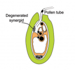 During the development of the plant reproductive lineages – the germlines – typically, single sporophytic (somatic) cells in the flower become committed to undergo meiosis. Here, Grossniklaus and colleagues review recent studies examining the molecular mechanisms underlying cell specification and the acquisition of reproductive fate in sexual and asexual plant species. See the Review on p. 229
Establishing neural crest identity: a gene regulatory recipe
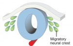 The neural crest is a cell population that contributes to a variety of derivatives, including sensory and autonomic ganglia, cartilage and bone of the face and pigment cells of the skin. Simões-Costa and Bronner examine neural crest development from a gene regulatory perspective and discuss how the underlying genetic circuitry results in the features that define this unique population. See the Review on p. 242
 (No Ratings Yet)
Get involved
Create an account or log in to post your story on the Node.
Sign up for emails
Subscribe to our mailing lists.