On how odd critters can answer important questions
Posted by Kim Cooper, on 27 March 2013
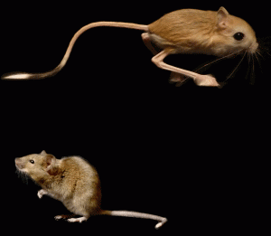
 Sproing! Sproing! Sproing! If there is one animal that deserves its own cartoon sound, it is the jerboa – a bipedal desert rodent with extraordinarily elongated hindlegs, fused foot bones, and loss of the first and fifth toes. I blogged here from China last spring during the most recent field collection of jerboa embryos, and now I’m excited to share news of the first research article using jerboas to answer a fundamental question in cell biology and skeletal growth and evolution: how do growth plate chondrocytes enlarge, and how do growth plates adjust cell size to contribute to differences in rates of skeletal growth? I have been derelict about entries, not sure what people would find interesting to read, so there is a lot of backstory that I will try to summarize. If anyone wants to see some of this story fleshed out, I would be happy to take blog post requests :)
I decided to postdoc with Cliff Tabin because of my interest in the limb as a structure to study morphological diversity, but “I am interested in limb diversity” isn’t a focused way to start a project. We spent a few months tossing around ideas for a specific animal to study wherein I laughingly proposed dinosaur, dolphin, and horse. That’s one great thing about Cliff – he took each of my crackhead ideas seriously and never said “no”. That meant my joking suggestion of horses turned to “Here’s your horse” when a student at Harvard introduced us to the jerboas. The jerboa and horse have converged on similar adaptations including loss of toes and fusion and elongation of remaining elements. I am actually starting to address convergence in horses as well since it turns out horse embryology isn’t impossible. But that’s a story for another day.
The early years were focused on acquiring specimens involving multiple trips to China to collect pregnant females and harvest the embryos. The benefit of going to China is that the species there hibernate through the winter and breed almost synchronously in the spring. This means that if we hit the timing right, we can get tons of embryos from a short collection. Unfortunately it takes some trial and error to get that timing right. When you work on a seasonal animal that means it can take several frustrating years to achieve success.
Meanwhile I embarked on an adventure to get the first research colony of jerboas established, and everything I’d read online said they would not take care of their offspring in captivity. It was the tiger mauling at the San Francisco Zoo in 2007 that brought to my attention the Association of Zoos and Aquariums and, more importantly, the International Species Information System. I sent an email to ISIS to ask if they had a husbandry manual or holdings records for any species of jerboa, and they responded with no husbandry manual but with the contact information for 11 zoos and conservation centers that had jerboas in their records at some point since the 1960s. I emailed all of them and got one amazing response from the Breeding Centre for Endangered Arabian Wildlife in Sharjah, UAE. This species of jerboa isn’t endangered, but that whole story and how I learned to raise jerboas is probably also best saved for another entry or this will get ridiculously long.
I started investigating mechanisms of digit loss and mechanisms of rapid skeletal elongation in parallel, but the embryos to study digit loss were in short supply until this last (amazing) collection. Meanwhile, the colony started to breed, and I realized that the significant time window for increased hindlimb elongation was in early postnatal stages – easy to get from my burgeoning colony without decimating the breeding population. I started with an analysis of BrdU labeling index…nothing exciting there. One day as I was looking at sections that were stained with H&E it struck me: those hypertrophic chondrocytes in the jerboa foot are HUGE.
So I dug through the literature and found a paper by Norm Wilsman and colleagues in 1996 that quantified the percent contribution of a number of factors to the daily rate of skeletal elongation (growth in microns per day). They determined that the process of volume enlargement during hypertrophy of the terminally differentiated chondrocytes contributes most significantly to skeletal elongation. Additionally, the size of those hypertrophic chondrocytes varied most between skeletal elements that elongate at different rates within an animal (ie the fast proximal tibia growth plate versus the slow proximal radius).
Hypertrophic chondrocyte size contributes significantly to skeletal growth and to differences in rates of growth, so how is cell size regulated? But that question turns out to be the second question in the pipeline. The first question was “How do these cells get big?” There was some discussion in the literature, primarily from Joseph Buckwalter and Peter Bush, suggesting that chondrocytes may enlarge by cell swelling – a disproportionate increase in cytoplasmic fluid volume. However when I talked to a couple of cell biologists at Harvard, I was met with some resistance. Plant cells swell, but they also have cell walls to contain the pressure. Animal cells don’t swell. If the neurons in your brain swell by as little as 10%, they rupture which causes some serious problems. Regulated volume increase and regulated volume decrease are so important that our bodies have developed mechanisms to stabilize cell volume in cases of shifts in blood plasma osmolarity. But hypertrophic chondrocytes looked really empty in transmission electron micrographs…
I decided to address this question of whether chondrocytes swell and put together a K99 proposal (which missed the payline by one point in the only submission I could make before hitting the 5 year mark). In the lead up to grant submission, Cliff put me in touch with Tim Mitchison in Systems Biology who introduced me to Seungeun Oh in Marc Kirschner’s lab. Seungeun had just finished her PhD in Spectroscopy at MIT and had joined Marc’s lab to apply microscopy methods to questions of cell volume control in the cell cycle. She had the perfect methodology to answer my question – using the retardation of a wavelength of light to quantify cellular dry mass contents. The very first experiment we did was on her old set up at MIT the day before my grant deadline to provide proof of principle. I had dissociated postnatal day 5 mouse tibia chondrocytes, and we carried the dish on the M2 shuttle across town to the basement of MIT. (Isn’t all of MIT a long basement corridor?) One of the first images we took was a perfect cluster of 3 cells of varying sizes showing a small cell, an intermediate cell that was more red on the phase shift heat map, and a third cell that was enormous and less red on the phase shift heat map indicating it hadn’t increased in total mass as would be expected for a larger cell maintaining high density. Even though we still had to collect data from many more samples and show that the cells remained spherical and didn’t just flatten out (which could also decrease the phase delay), we were so excited and gave each other a good high five. That image from the very first day became panel “a” of Figure 1, because we never again saw such a perfect cluster of cells together demonstrating the effect of cell swelling on phase shift in a single image. It’s like they were waving at us with a taunting “You’re right!”
That first day led to 2 ½ years together in a small dark room imaging cells, countless hours of clicking on images in Matlab, writing, re-writing, reworking figures, and discussions of osmolarity and properties of light in the hybrid language of an organismal biologist and a physicist. It was a perfect merging of two disciplines. From that initial observation in the mouse proximal tibia, we went on to discover that there are three phases of chondrocyte enlargement: an initial phase where chondrocytes increase in volume while maintaining a “normal” dry mass density of about 18%, a second phase where cells swell and dilute their dry mass to about 7%, and a final phase where cells continue to get larger but maintain low dry mass density. Larger cells get bigger by extending the third phase, and smaller cells truncate the process that’s shared by all chondrocytes observed in this study. To get at a genetic mechanism controlling this process, we investigated the cell phenotype in animals that were conditional mutants for insulin-like growth factor 1. We found that not only do the growth plate specific differences in cell size disappear, but that igf1 controls the third phase that is most variable in growth plates elongating at different rates. This puts the igf1 pathway in a perfect place to investigate the genetics underlying evolutionary differences in growth rate. Stay tuned.
But for all of the answers this paper provided, it opened a whole new can of question mark shaped worms. How do animals establish different skeletal proportions? (The original question I set out to answer.) How does the cell carry out metabolic functions in the face of a 3-fold decrease in dry mass density? How does the cell regulate its membrane surface area? And a big one – how in the heck do these cells drive all that water in to dilute the cytoplasm? As I’m preparing to set off and establish my own lab, I’m looking forward to where these questions and more will lead.


 (6 votes)
Awesome post. I’m a big fan of cool critters. So, where to now?
quite a dream come true research.. inspiring.
Terrific post.
I can’t help but wonder how many experiments are carefully tucked away on M2 shuttle passengers. They all look so calm with their ipods. To think that some of them may be carrying hypertrophic chondrocytes of jerboas for wavelength retardation analyses….
Of course I had all of the appropriate transport paperwork allowing me to do that… I was marveling on the Red Line to Kendall yesterday how strange it’s going to be to live in a city where it’s not completely normal for other random passengers to be discussing their IRBs.
Also, J Gross is coming for EB/AAA – will you be here too?
Nice to see the wrap-up from your previous China travel posts/blog. Does this mean no more posts from Xinjiang? Ir perhaps you are considering a position at Qinghua Daxue….