Vote for your favourite image from the MBL Embryology course
Posted by the Node, on 19 August 2025
We are delighted to bring you the return of our image competition in collaboration with the MBL Embryology course at Woods Hole. We’d like you to vote for your favourite image from the stunning submissions from the students that attended the 2025 course. The winning image will be published on the front cover of Development later this year.
Please vote for your favourite image using the poll at the bottom of the page. The voting will close on Wednesday 3 September.
Thank you and good luck to the following researchers for their contributions:
Virginia Panara, Shirley Ee Shan Liau, Sonoko Mizuno, Ignacio Casanova-Maldonado, Max Makem, Johnny Vertiz, Arthur Boutillon, Anthony Wokasch, Aria Zheyuan Huang, Amartya Tashi Mitra, Nathanial Sweet, Paul Maier, Shivangi Pandey, Marie Lebel, Chloe Kuebler, Nicole Roos
Browse through the gallery (click to view full image)
Sonoko Mizumo, Virginia Panara, Shirley Ee Shan Liau
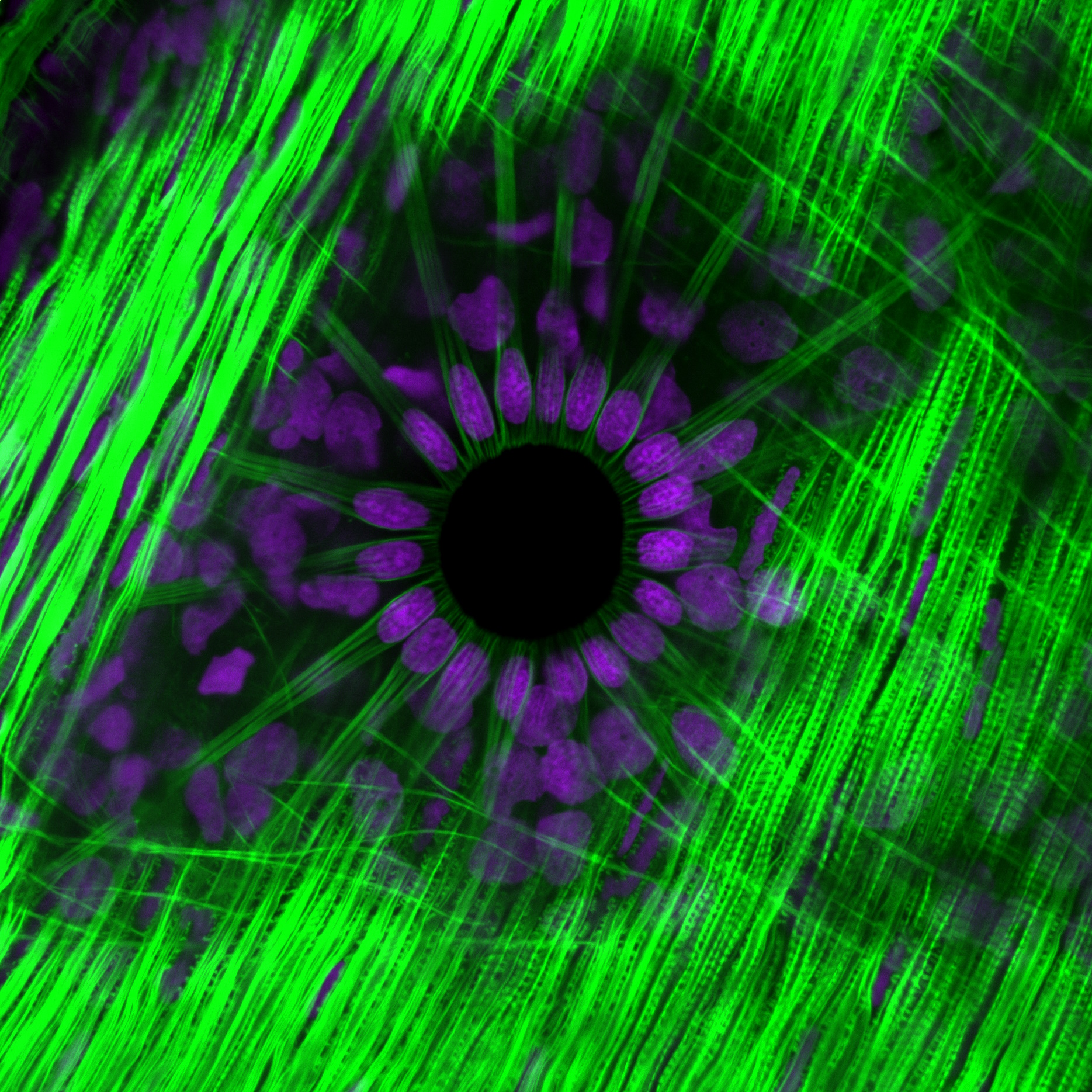
Fluorescent imaging of the muscle ring surrounding a Longfin Squid chromatophore
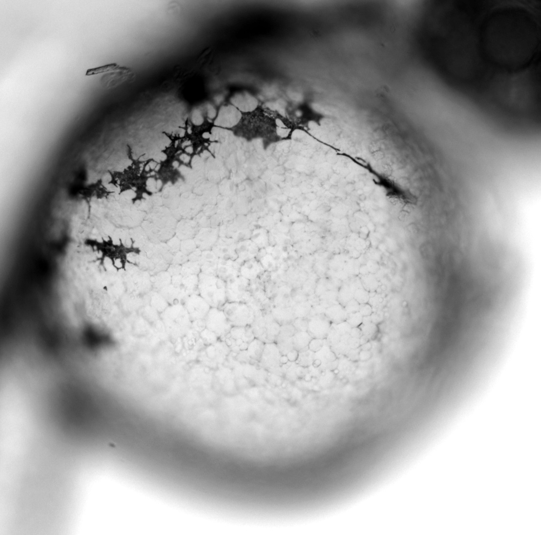
Sonoko Mizuno
Bright field imaging of melanocytes in a Zebrafish larva
Ignacio Casanova-Maldonado, Max Makem & Johnny Vertiz
Mitochondria (Magenta), Cell membrane (Cyan). Olympus FV4000 Confocal microscope, 10X (N.A: 0,4).
Ignacio Casanova-Maldonado, Max Makem & Johnny Vertiz
Nuclei (Cyan), Actin (Red). Olympus FV4000 Confocal microscope, 4X (N.A: 0.8)
Ignacio Casanova-Maldonado, Max Makem & Johnny Vertiz
Actin (magenta), circulatory system (red) mitochondria (green) and nuclei (cyan). Olympus FV4000 Confocal microscope, 10X (N.A: 0,4)
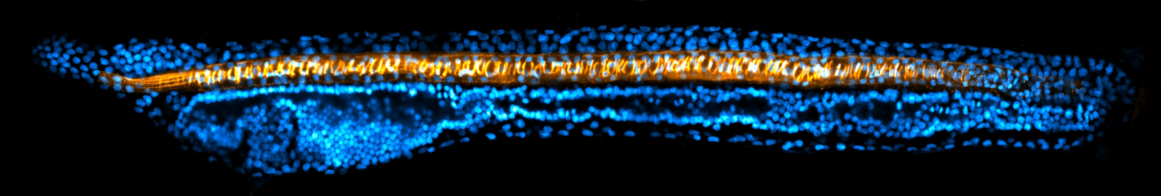
Arthur Boutillon
Amphioxus larva (stage L1) stained for nuclei (DAPI, blue) and phosphorylated myosin II (orange), imaged by point scanning confocal microscopy and prossessed using ImageJ.
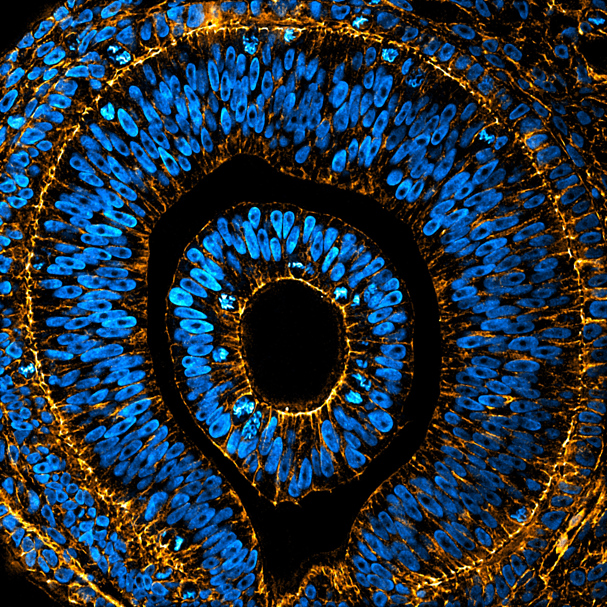
Arthur Boutillon
Embryonic eye of an Anole lizard stained for nuclei (DAPI, blue) and F-actin (Phalloidin, orange), imaged by spinning disc confocal microscopy and prossessed using ImageJ.
Arthur Boutillon
Wing disc of the butterfly Vanessa cardui stained for F-actin (SiR-Actin, orange) and membrane (PKmem555, magenta), imaged by point scanning confocal microscopy and prossessed using ImageJ.
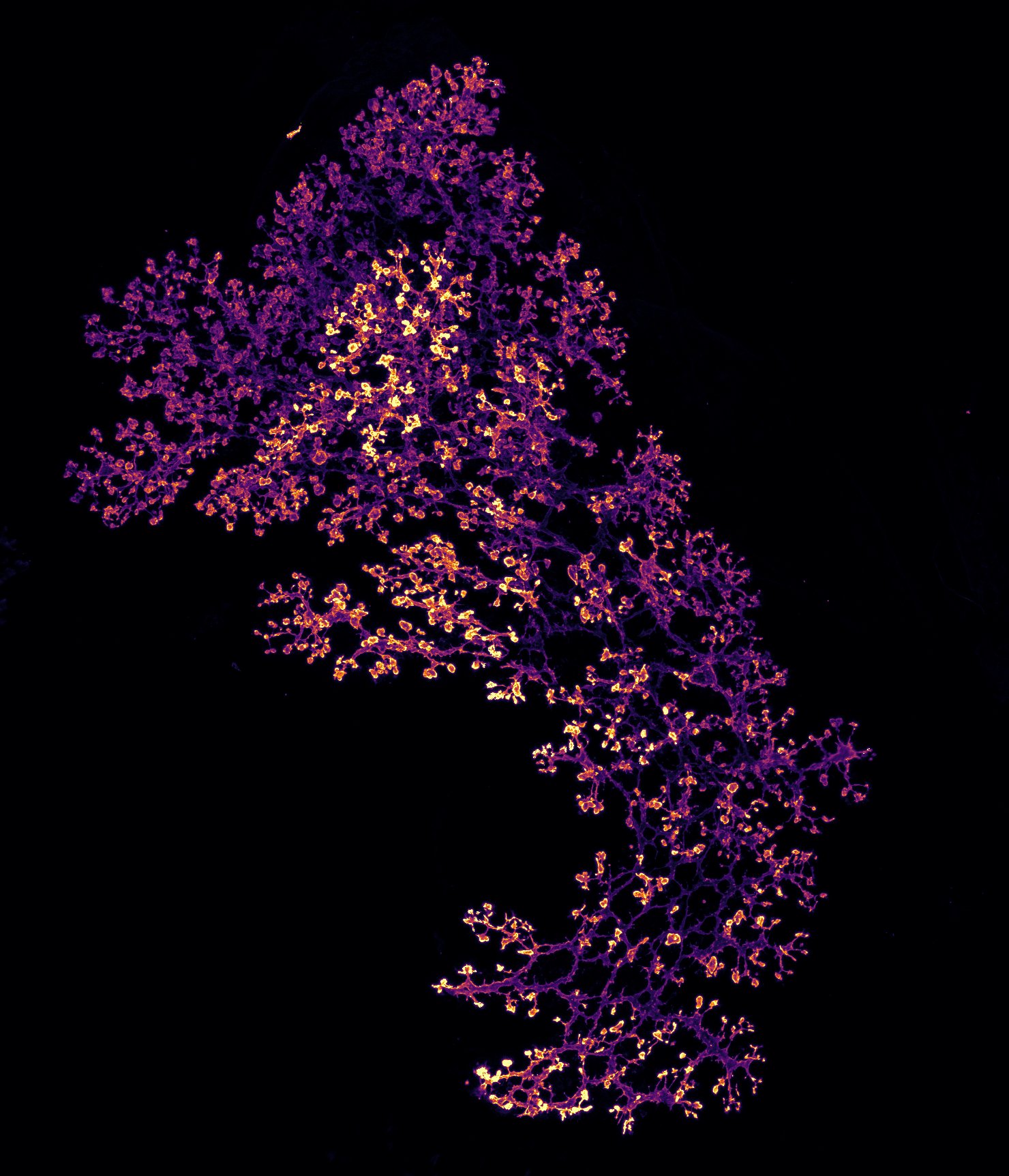
Anthony Wokasch
Ductal branching of an E15.5 mouse pancreas. E15.5 whole-mount pancreas labeled with Mucin-1. Imaged on the Olympus FV4000 Confocal Laser Scanning Microscope (20X) and processed using FIJI Max projection.
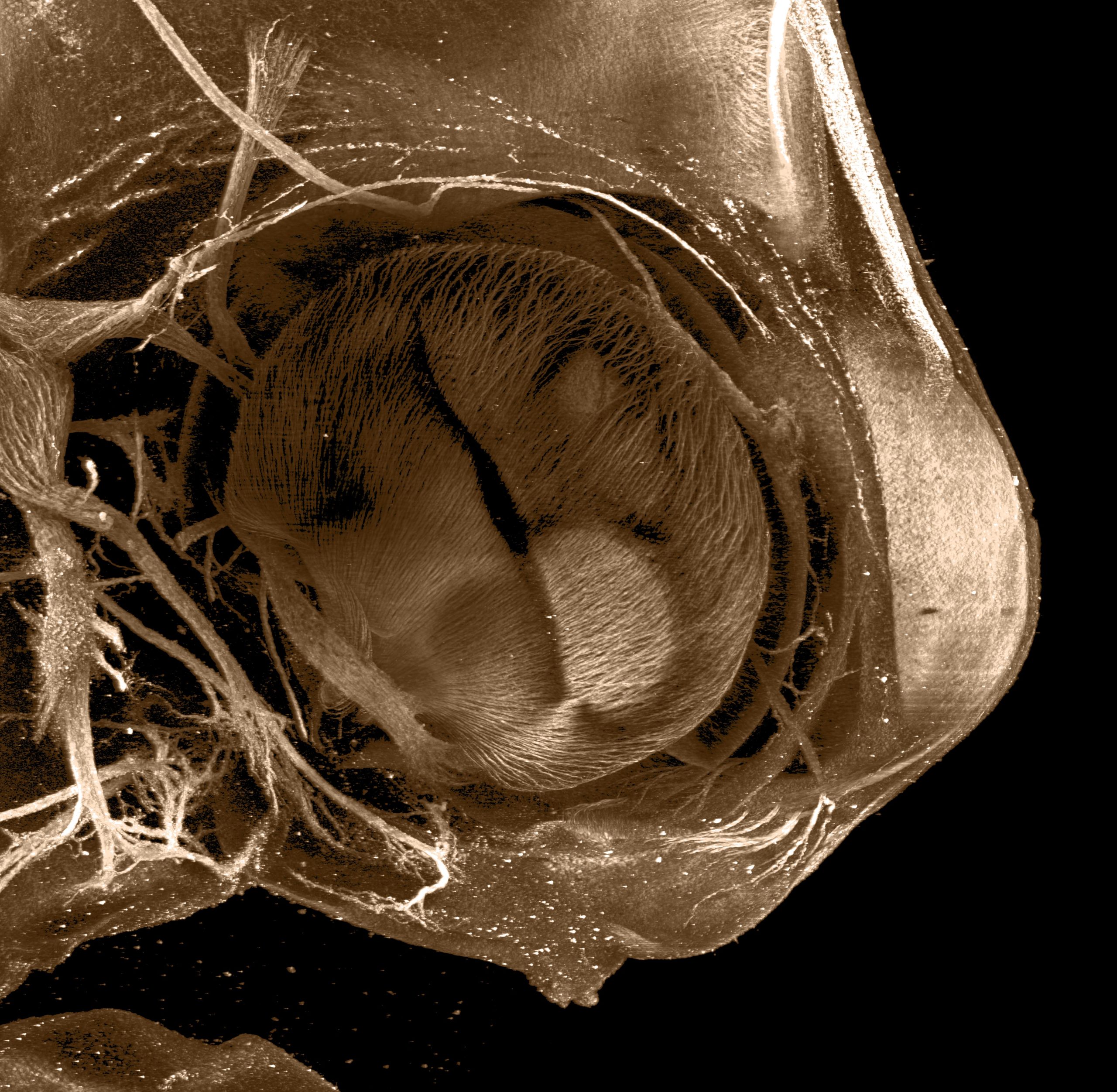
Anthony Wokasch, Aria Zheyuan Huang, Amartya Tashi Mitra, Nathanial Sweet, Paul Maier
Close-up of a Red eared slider turtle (Stage 14), Trachemys scripta, optically cleared and stained with acetylated tubulin. Imaged on the LifeCanvas MegaSPIM Light Sheet.
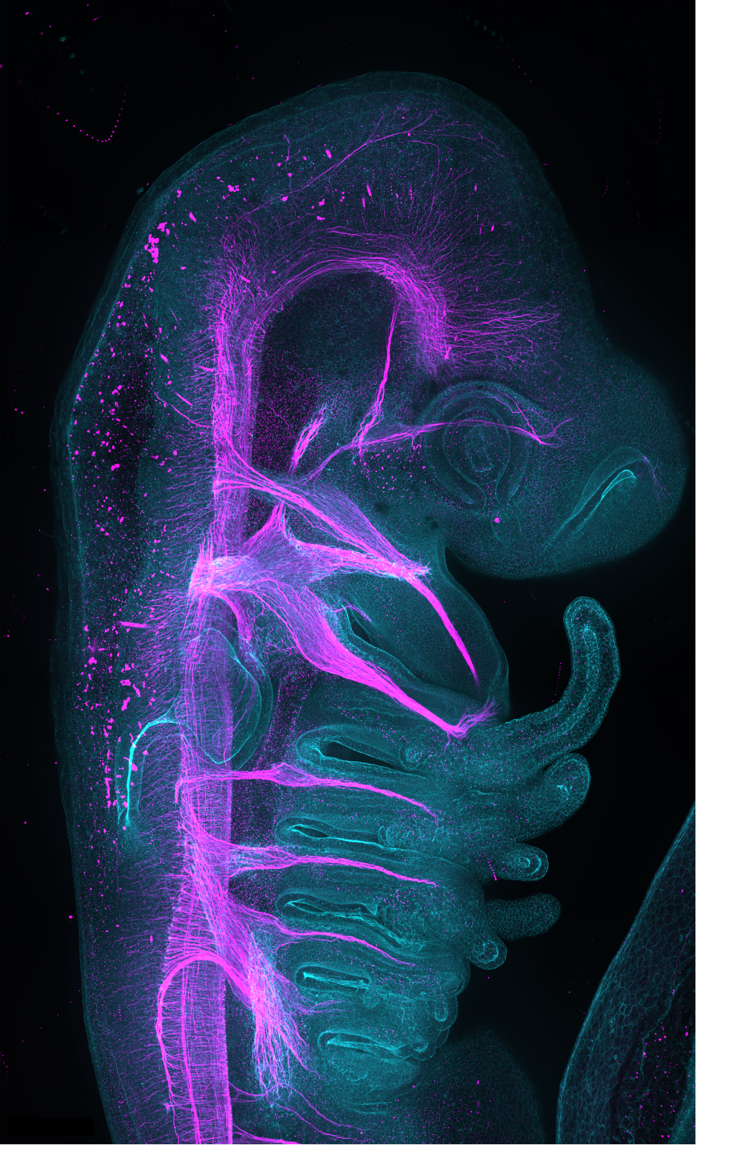
Amartya Tashi Mitra, Aria Zheyuan Huang, Nathaniel Sweet, Anthony Wokasch, Paul Maier
Red-eared slider (Trachemys scripta) embryo stained with acetyl-alpha tubulin, labelling neurons. Optically cleared and imaged on LifeCanvas technologies MegaSPIM light sheet microscope.
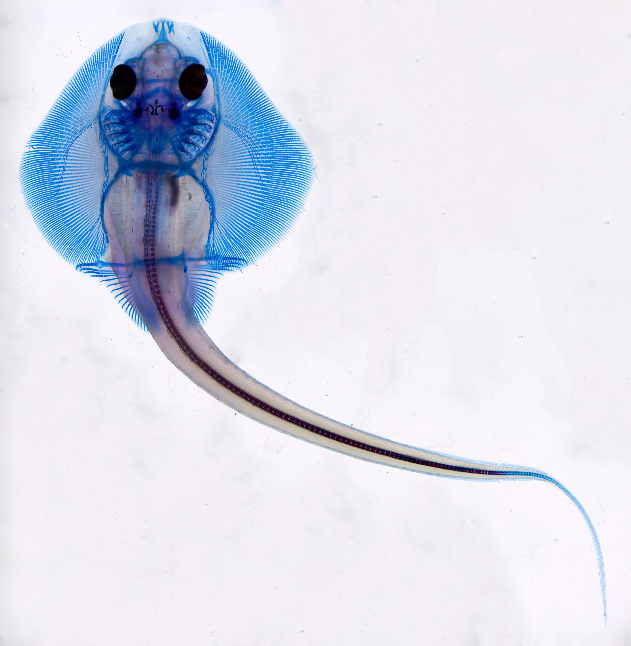
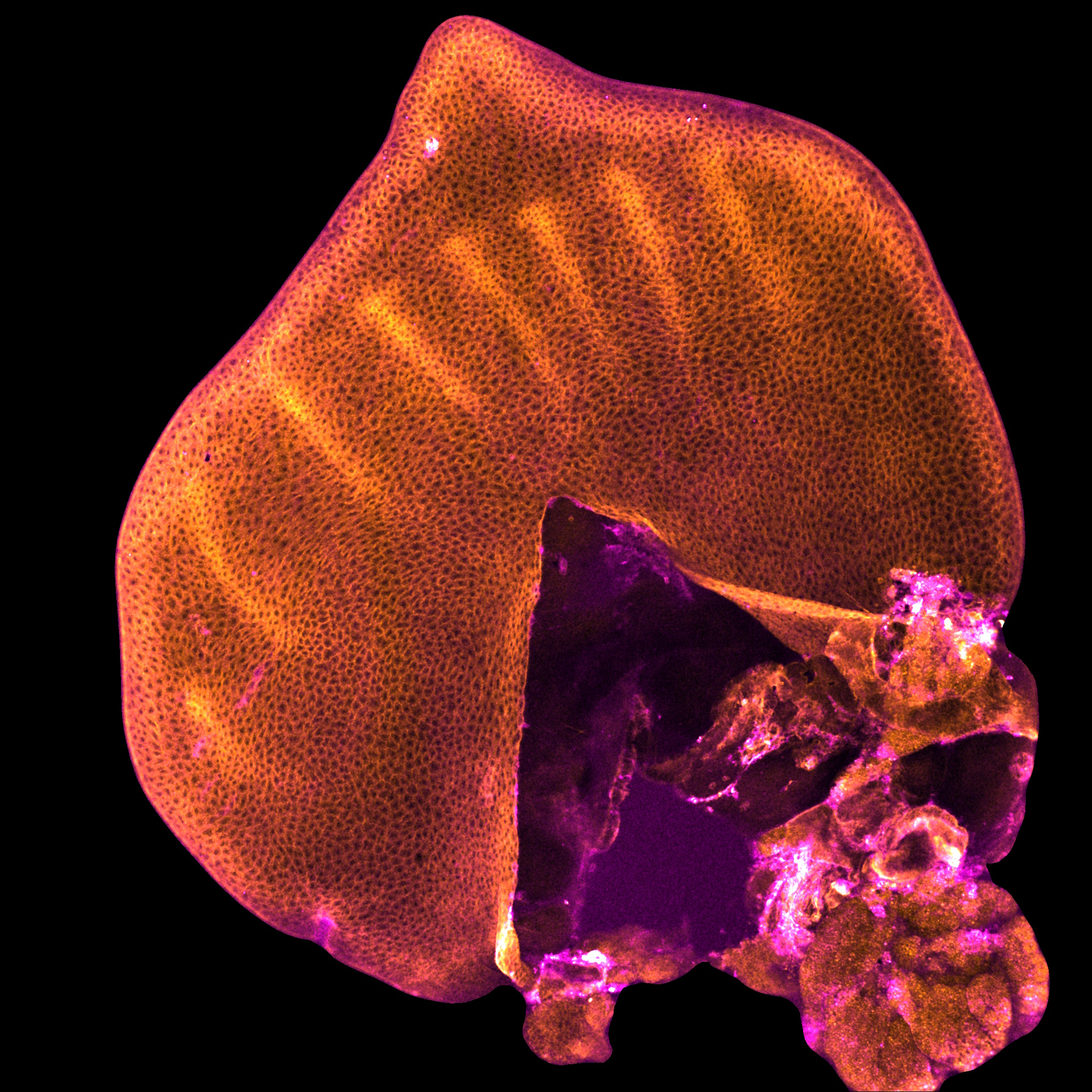
Amartya Tashi Mitra
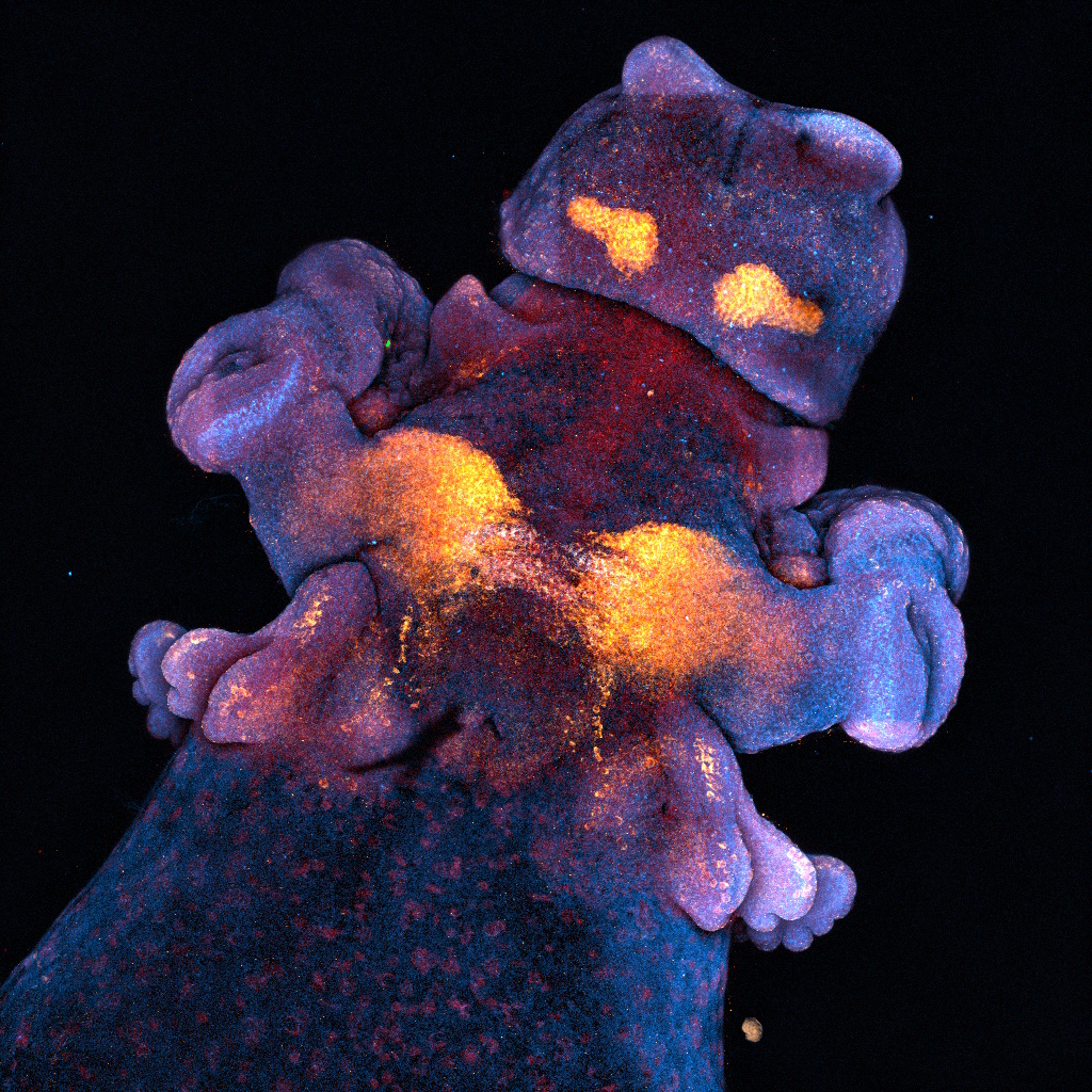
Skeletal preparation of little skate (Leucoraja erinacea) embryo labelling cartilaginous tissue in blue and calcified tissue in red. Imaged on Leica MZ10F stereomicroscope, assembled using focus stacking and tile stitching.
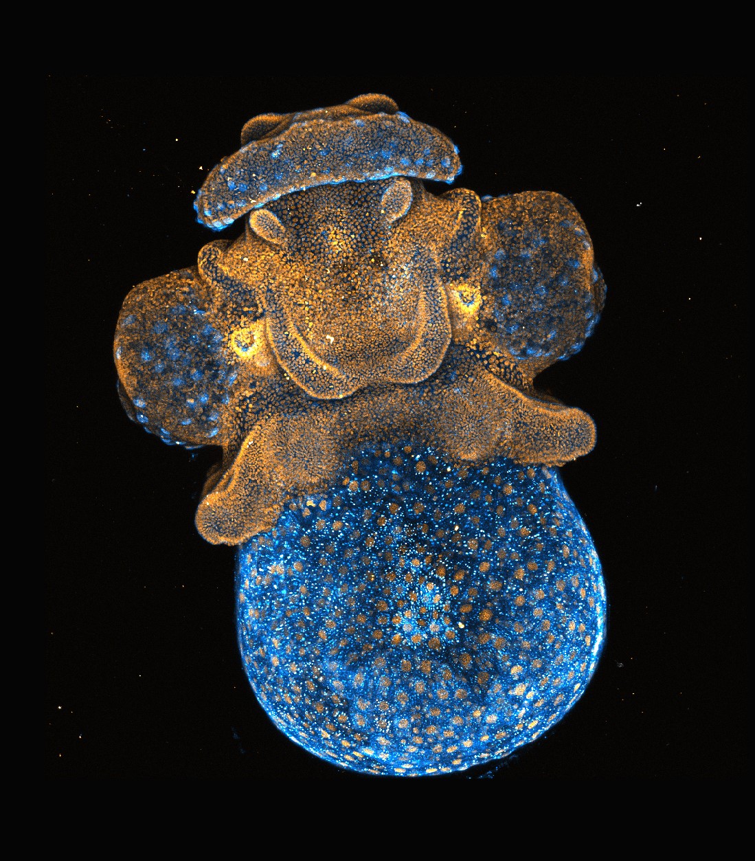
Amartya Tashi Mitra
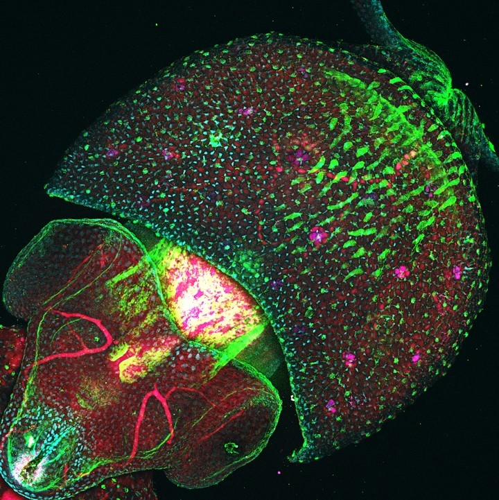
Longfin inshore squid (Doryteuthis pealei) embryo with plasma membrane (CellMask Green) and nuclear (Hoechst) labelling in blue and orange respectively. Imaged on Nikon AXR laser scanning microscope.
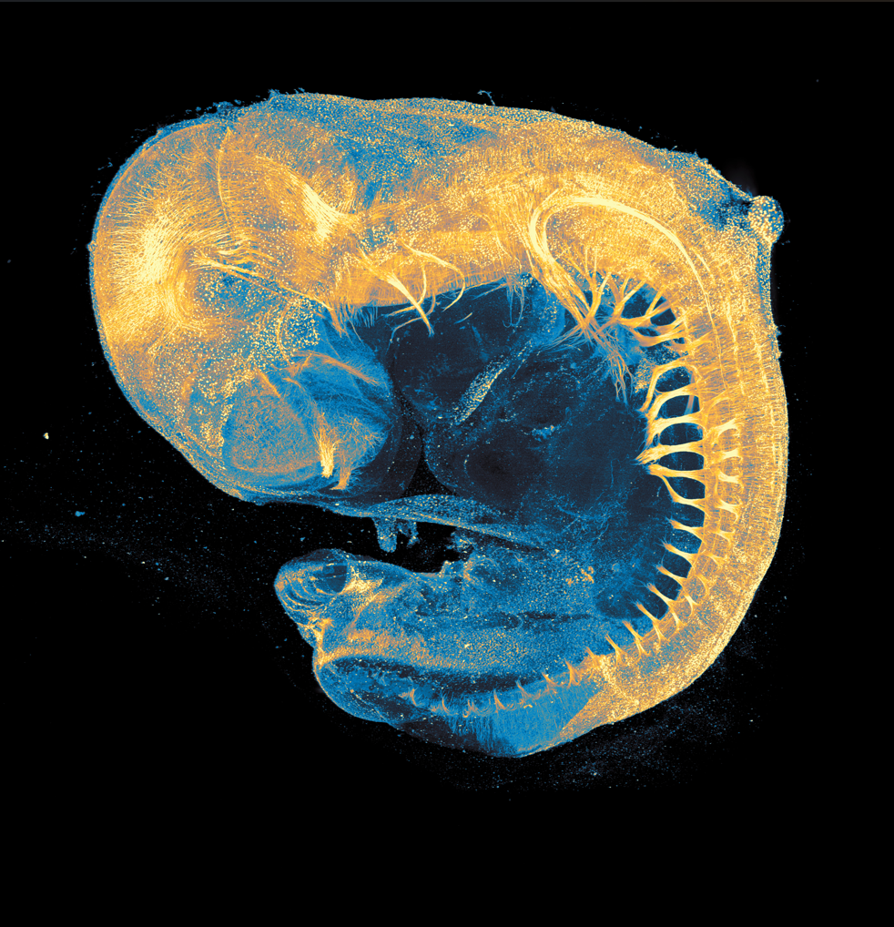
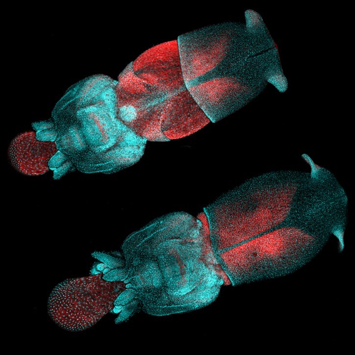
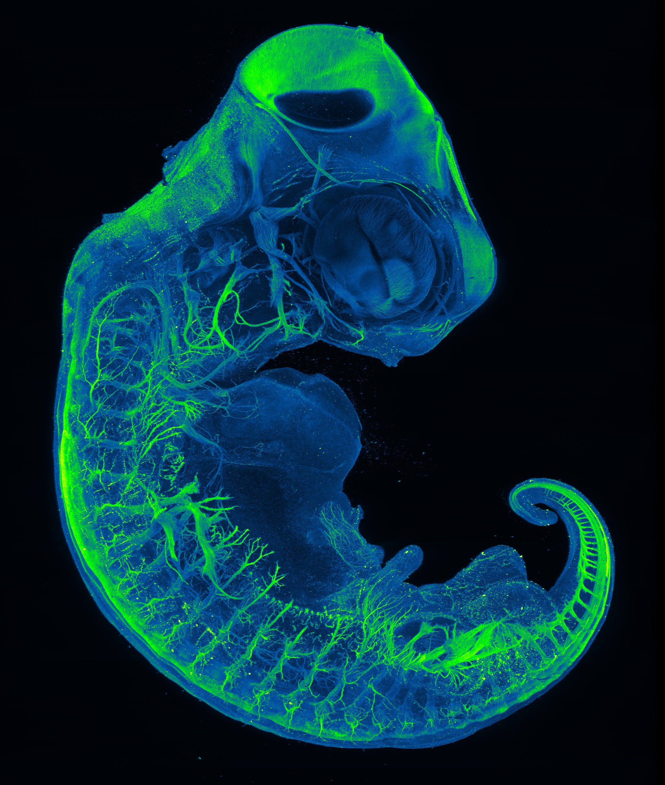
Aria Zheyuan Huang, Amartya Tashi Mitra, Nathanial Sweet, Anthony Wokasch, Paul Maier
CD-1 mouse embryo at embryonic day 10.5, optically cleared and stained with acetylated tubulin (yellow), imaged on a LifeCanvas MegaSPIM Light Sheet.
Shivangi Pandey
24hpf Zebrafish embryo stained for Acetylated tubulin (red), Prox1(green) and DAPI(blue). The image was acquired using a Yokogawa W1 (Eclipse) Spinning Disk microscope. The image was processed using ImageJ.
Shirley Ee Shan Liau and Shivangi Pandey
An early-stage skate embryo stained for DAPI (cyan) and Tuj1 (Magenta). The image was acquired using a Yokogawa W1 (Eclipse) Spinning Disk microscope. The image was processed using ImageJ.
Marie Lebel, Shivangi Pandey
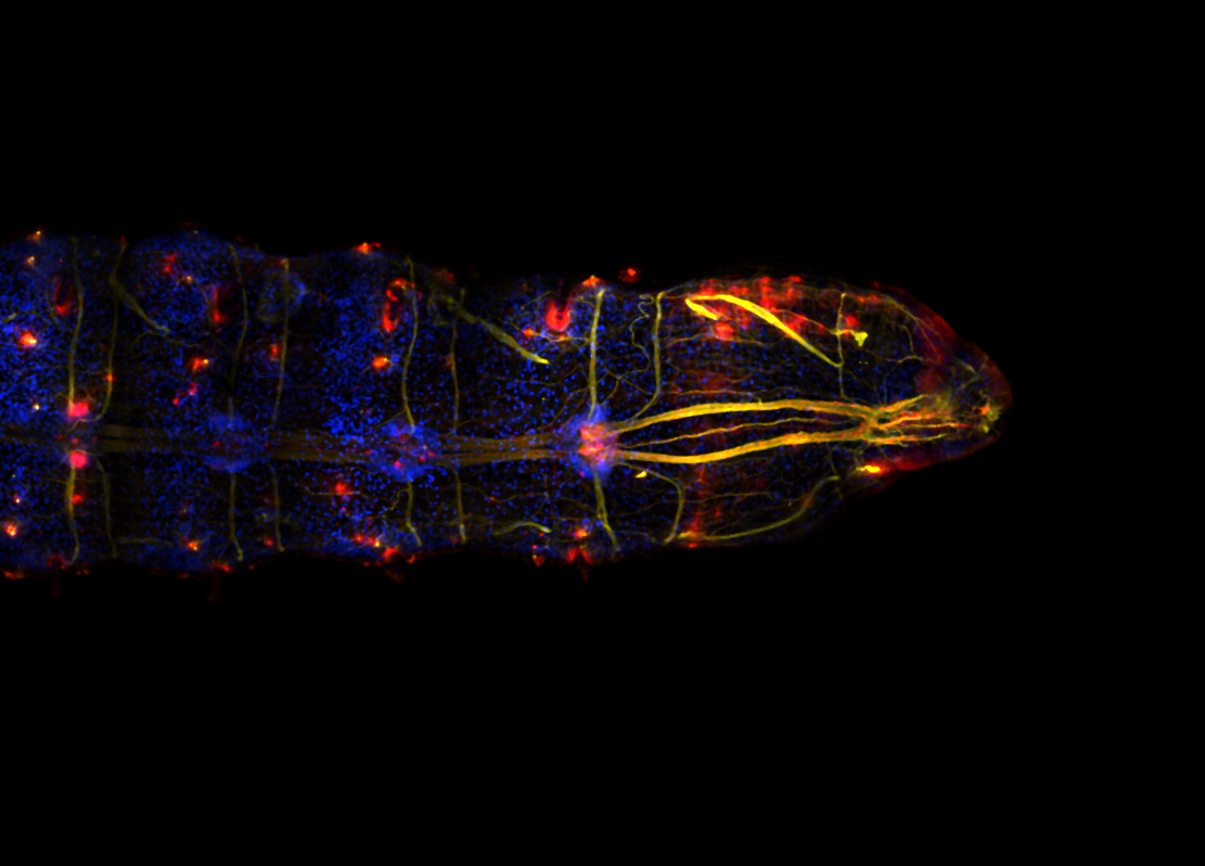
Regenerating posterior end of a Capitella teleta juvenile seen from the ventral side, 3 days post amputation imaged with a scanning confocal microscope (Nikon AXR NSPARC; 20x, NA 0.8 objective). Nuclei are in blue, neurons in yellow, and serotonergic neurons in red.
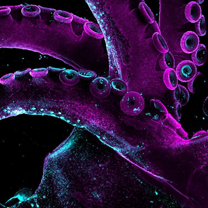
Marie Lebel, Shivangi Pandey
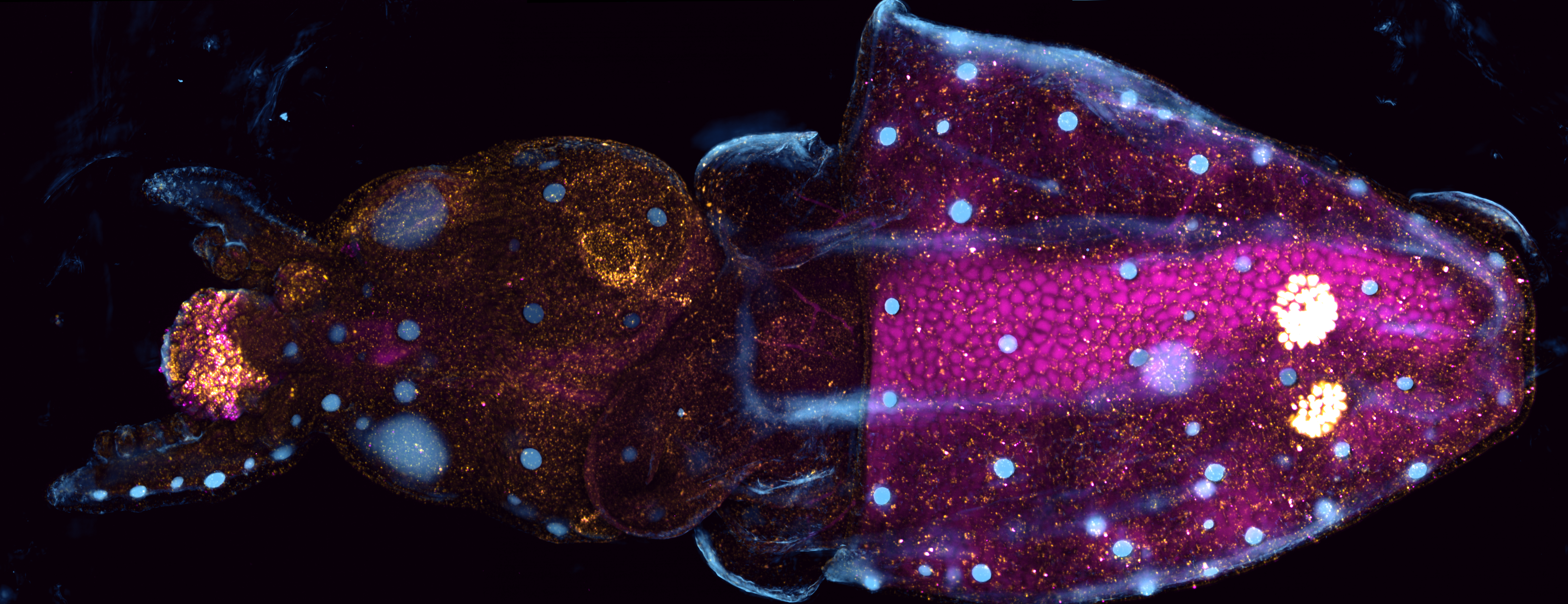
Late squid embryo, with a tentacle amputated 3 days prior, imaged with a spinning disk confocal microscope ( Andor BC43; 10x, NA 0.45 objective) . TRITC (yellow) and CFSE (magenta) were injected in the vasculature a day before amputation. The cyan signal corresponds to the inverted brightfield, highlighting the eyes and chromatophores.
Amartya Tashi Mitra, Chloe Kuebler, Shirley Ee Shan Liau
Longfin inshore squid (Doryteuthis pealei) embryo HCR in-situ. mRNAs for elav, optix, and pcdh17 mRNAs represented in red, blue and orange respectively. Imaged on Olympus FV4000 laser scanning microscope.
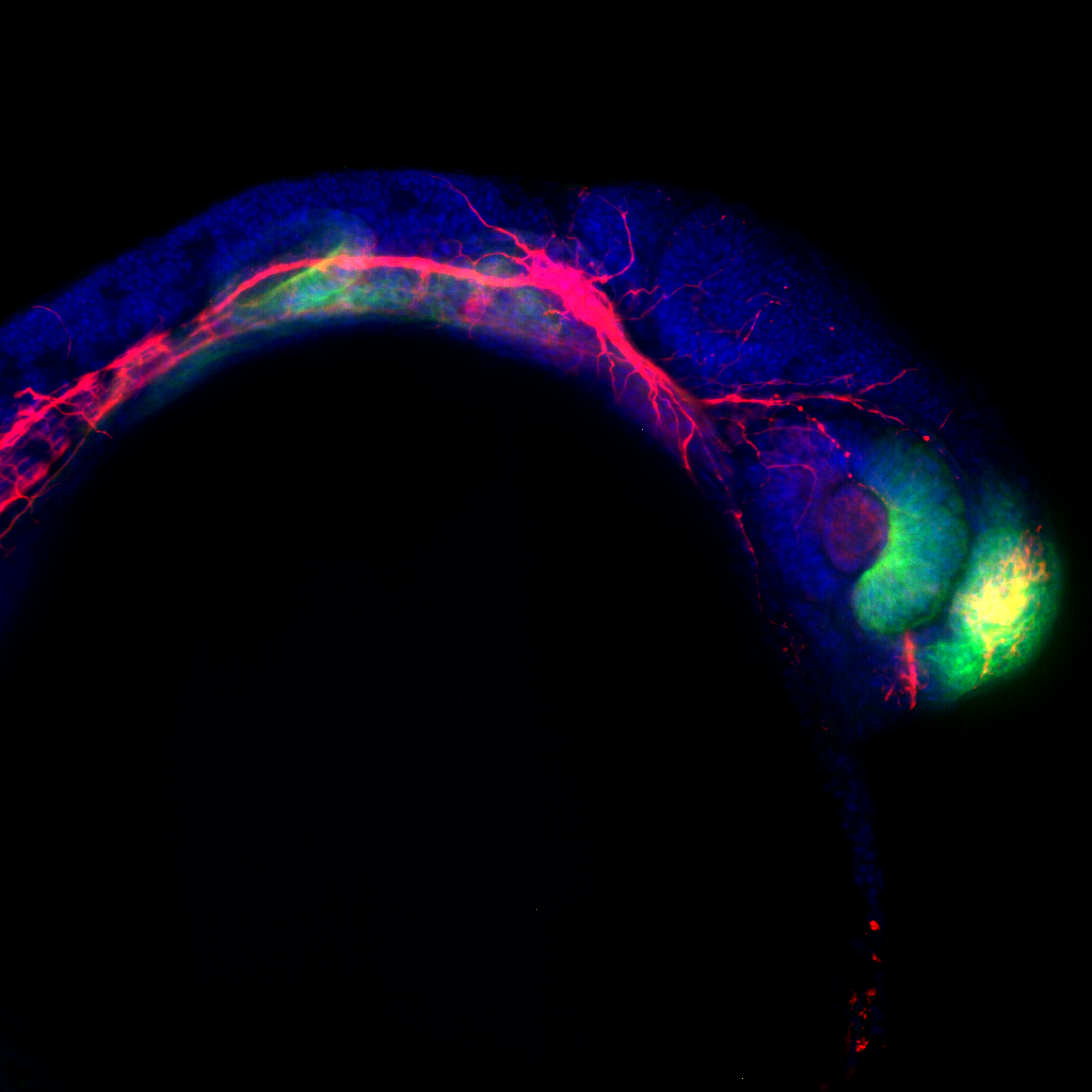
Nicole Roos and Anthony Wokasch
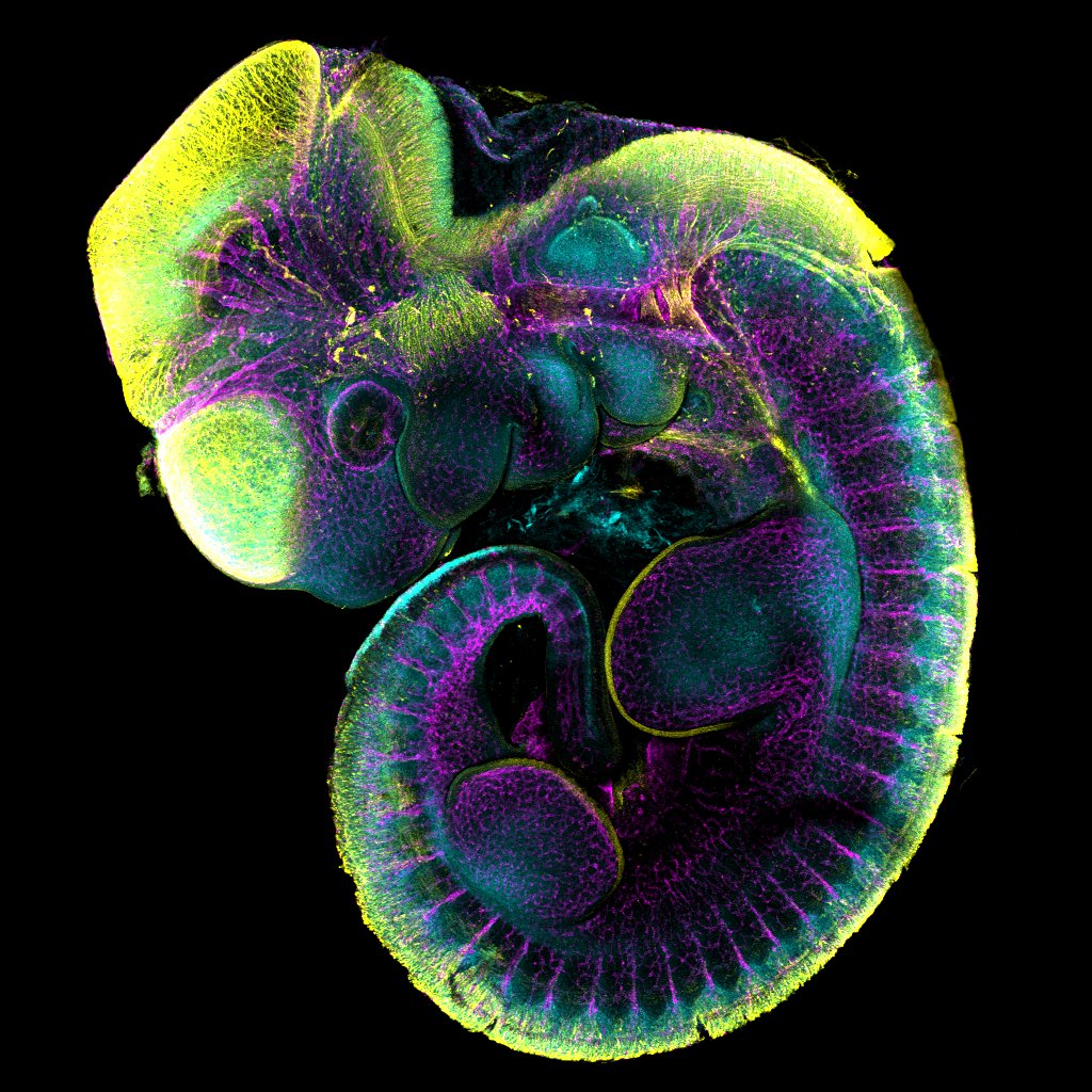
Mouse E10.5 embryo immunofluorescent staining of Sox9 (cyan), alpha-tubulin (yellow), and endomucin (magenta) protein. Image captured on Evident FV4000 point scanning confocal, lens UPLXAPO4X, na = 0.16, zoom = 1.04. Image processing conducted on Fiji.
Pick your favourite image
 (193 votes)
22 thoughts on “Vote for your favourite image from the MBL Embryology course”
Leave a Reply
Get involved
Create an account or log in to post your story on the Node.
Sign up for emails
Subscribe to our mailing lists.
Read the latest Development issue
Most-read posts in May
- From comparison to mechanism: decoding heart regeneration
- A day in the life of a Reviews Editor (at Development)
- What does a Reviews Editor do?
- New evo-devo textbook ‘Eco-Evo-Devo: The Environmental Regulation of Development, Evolution, and Health’
- preLighters’ choice – A curated selection of recent preprints
Curled up, pondering on existence
An excellent work with beautiful and clear light effects, reflecting the Artist’s in-depth vision and clarity of thought.
Spiral of birth !
They are all beautiful–hard to pick a favorite.
Love the intricacies of the neural network!
Such beautiful definition and shape!
The images are awesome 👍🏻
I wish we could choose more than one. Maybe rank voting? There are so many stunning images.
Sick pic
That’s an amazing image!
This is amazing!
What a amazing image love to see how you created that
Wish more could be selected
They are eall so beautifull
Nature never ceases to amaze
Amazing photos! Shows the divine hand of nature!
Every entry is stunning.. best wishes for the brilliant young scientists!!
These are amazing . Vegetable Creator has made all things beautiful.
Love the details and colors
Good job guys
Goes to enforce the thought that ‘all’ life has fascinating form , often our focus being on the human kind ….
Wow! I am so impressed!
Wow! This is amazing!
I am astonished at the beauty of these images. I am not a scientist so have never seen such embryos like this. It was so hard to pick, all stunning. Rather mind altering.
Fascinating to see these images in their embryonic state.
Beautiful images! Hard to pick a winner . Congrats to all!