

Going with the flow and the art of redirection
Posted by Hoang Anh Le, on 4 November 2026
Hoang Anh Le
Research fellow in Cell and Developmental Biology, University College London
TLDR: Approach science with the mindset of a waterbender, be like the flow of a river, flexible, malleable, adapting, and most importantly, enjoy the ride and stay curious!
The philosophy of waterbending
Ok, I know you probably think I’m a bit wacky with that opening heading, but bear with me! There is this great cartoon series by Nickelodeon from the early 2000s that I grew up watching (and rewatching still) called Avatar: The Last Airbender. The show brings you into a world of four nations, where people from each nation can manipulate one of the four elements: water, earth, fire, and air through a type of martial art called bending. The show’s premise isn’t important for this piece, but one of the greatest things about it is the philosophy behind each bending form, and for me, the best has always been waterbending. “Water is the element of change.” Waterbenders “let their defence become their offense, turning their opponents’ own forces against them. The people of the Water Tribes are capable of adapting to many things.” As much as one might think this is just a kid show, this philosophy has stuck with me and underlies much of how I approach science.

After finishing my PhD in cancer migration and metastasis in 2020 at the Cancer Research UK Scotland Institute, I wanted change. I wanted to explore more complex systems than cell culture. I became interested in how cells respond to mechanical forces, particularly how immune cells integrate these signals to regulate a process called macropinocytosis, also known as cell drinking. I’m not sure where this fascination with immune cells came from, because before this, I had never worked with them. But the idea of a migratory cell type capable of morphing, branching, and constantly gulping up stuff piqued my interest and, honestly, sounded like a fascinating system to explore. I’d known of Roberto’s work for a while and knew his expertise in in vivo mechanobiology, so I approached him and proposed a project to look at the mechanobiology of early macrophages (also known as embryonic myeloid (EM) cells) in vivo and determine how substrate stiffness regulates macropinocytosis using Xenopus embryos. I applied for the Sir Henry Wellcome fellowship and was lucky enough to get funded. This marked my biggest career transition, moving from relatively neat, clean cultured cell lines to frog embryos. It was also daunting because, unlike transparent zebrafish, frog embryos remain a rather unusual model for immunobiology, and this project was about to be led by someone who had no prior experience working with live embryos. But just like the philosophy of waterbending, one can either look at changes as something to fear, or they can look at them as new opportunities for growth and adaptation, turning fear into excitement and curiosity.
The first goal
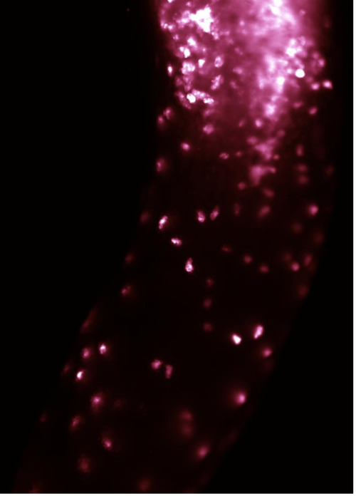
The first major goal of this project was to establish an ex vivo explant system of myeloid cells. If imaging through a Xenopus embryo is difficult because of yolk platelets present in every cell, then these same yolk platelets also allow Xenopus tissue to be isolated and cultured independently for an extended period. An ex vivo explant would allow us to perform high-resolution imaging and cellular perturbations. I remember those first few months of constant trials and errors vividly. We had three main concerns at the time. First, isolated primary blood cells are cumbersome to culture in humans and mice, so we had no idea whether the same problem would apply to Xenopus myeloid cells. The second concern was how to establish a dissection and culturing method for a piece of tissue that nobody has ever worked on. Third, labelling these myeloid cells requires microinjecting into two rather small, obscured-looking blastomeres in 16- or 32-cell-stage embryos. Luckily for us, the last concern did not need to start from scratch, as some early work on microinjection and labelling was done by the Enrique Amaya lab at the University of Manchester1,2 (https://research.manchester.ac.uk/en/persons/enrique.amaya/). I cannot recall how many microinjections I did or how long it took me to comfortably recognise the right blastomeres, but I do remember the day I saw those early myeloid cells glowing in one of the injected embryos. I ran to Roberto’s office and told him he needed to come take a look. These cells are difficult to target, but you would know in a heartbeat if you had labelled them correctly because they spread out individually and colonise the entire embryo. They also look beautiful under the microscope with their characteristic branches that closely resemble those seen in zebrafish and mice.
Now that we got them labelled, the real challenge was to culture them. Roberto taught me how to do my first dissection. He was always so enthusiastic about teaching. I remember those days when it was just the two of us sitting in the embryology room until 8 or 9 PM, testing different ways to cleanly dissect out the anterior ventral blood island, the tissue region where the myeloid cells come from. The original idea was to culture just the myeloid cells. However, we quickly realised that because these cells migrate individually, it would be impossible to separate them one by one into a pure population, as is often done for collectively migrating cells like the neural crest. It was also not possible to remove the top tissue layer of the explant – the ectoderm – without affecting the myeloid cells below. To add to the complication, this ectoderm layer causes the dissected explant to curl up quickly into a ball, even if the culturing dish is coated with fibronectin. After quite a bit of brainstorming, I came up with the idea of gently placing a small piece of glass on top of the explants to prevent them from curling up and promote their adhesion to the substrate. To ensure I did not compress the tissue too heavily, I added a small amount of vacuum grease to the two opposite edges of the glass piece, and this essentially was how the protocol for culturing the anterior ventral blood island was born. I still have goosebumps reminiscing about the day when I collected the time-lapse images and saw the explants spread on the surface; then came the waves of myeloid cells radiating out from the centre. The first major milestone was achieved!
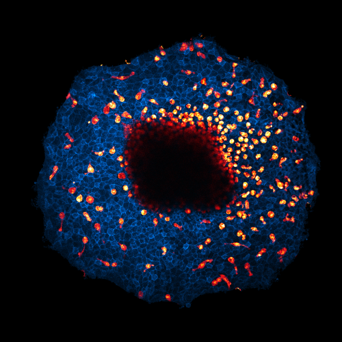
Ectodermal cells in blue (GFP-membrane), embryonic myeloid cells in red (fluorescence dextran).
The quest to control tissue flow
It wouldn’t make a good story without the drama because a new problem quickly arose. Since we could not separate the mesoderm from the ectoderm, our explants would permanently be a co-culture of at least two different cell populations. This makes it incredibly challenging to image macropinocytosis because the surrounding ectodermal cells would also take up the fluorescent dextran, making it difficult to differentiate the signal from the myeloid cells. Secondly, because of this ectodermal layer, it was also impossible to culture the explants on softer hydrogels since they could not attach and spread. This makes studying the role of substrate stiffness on myeloid cells’ macropinocytosis almost impossible. After months of unsuccessful attempts, the philosophy of waterbending came to mind again: redirection. What if the co-culture aspect of our explant system wasn’t so bad after all? I noticed that myeloid cells outside the explant display a rounded-up morphology with an erratic migration behaviour, unlike their more streamlined morphology inside the explant. This suggested that even if I could separate these myeloid cells, their natural biology would be severely altered, which was not the best-case scenario. At the same time, the startling radial migration pattern that these cells display also became hard to ignore. A spark flashed in my mind: what if the direct interactions between these myeloid cells and the ectodermal cells weren’t just mere touching? What if the co-culture system reflected how these cells interact in vivo? What if being unable to separate these cells apart was a blessing in disguise? We subsequently showed that our explant system indeed preserved the natural development and cell-cell interactions of myeloid cells. We decided to leave macropinocytosis behind and refocus on the physical interactions between myeloid cells and the surrounding ectoderm. What if movement of the ectoderm could explain the striking radial movement of myeloid cells? What if this “tissue flow” could act as a guidance signal that myeloid cells could follow, much like a river could guide a moving boat? This question is exciting because much of cell migration research has been done on isolated cells moving on a matrix-coated dish. While this approach has revealed a lot about the fundamental mechanisms of cell migration, it lacks the physiological relevance found in vivo because it ignores a fundamental fact that in vivo cells are rarely, if ever, alone. These cells are always surrounded by other cells, yet the effect of cell-cell interactions on migration is often underappreciated. After manually tracking hundreds of myeloid cells, I noticed that their movement often followed that of the surrounding ectodermal cells. When the ectoderm retracted, the myeloid cells would often retract their protrusions. When the ectoderm jiggled, the embedded myeloid cells would jiggle in the same way. If we disrupted the movement of the ectoderm, this also impacted the movement of the myeloid cells. This observation was later supported by a correlation analysis and modelling by our in-house computational biologist Jonas Hartmann and an external collaborator, physicist Ricard Alert. However, to directly establish a causal relationship between the movement of these two cell populations, we needed a way to control ectoderm movement at will. This turned out to be the hardest but most crucial question to answer.
The first idea was to create a flow of viscous liquid on top of isolated myeloid cells. The literature suggested using polyethene glycol (PEG), as this has been used to compress cells in culture3. However, Xenopus explants did not like PEG and would peel themselves off the dish. The thick and viscous liquid was also a nightmare to control. This was out of the window. The next idea was to stretch the explants cultured on a sheet of PDMS. But stretching generated tension and would cause the tissue to rip apart and detach. The stretching needed to be constant and continuous over time, which would require the automation and engineering we did not have the capacity or resources for at the time. Another idea bites the dust. Then I thought: “If explants are too difficult, why don’t I go back to my roots and try with cell culture? Afterall, I was great at cell culture!” What if we co-culture an epithelial-like cell line such as MDCK with a macrophage line such as RAW264.7, then scratch a wound in the monolayer and watch the MDCK cells flood in to fill the gap; perhaps then we would be able to observe the macrophages moving along? These cells are easy to grow, so this sounds like an easy experiment! Well… it was not! One thing I didn’t anticipate was that the junctions between MDCK cells and their substrate adhesions were incredibly strong, so as the monolayer grew, they pushed out all the co-cultured macrophages, which were much less adhesive! No matter what I did, I couldn’t get these macrophages to intercalate into the MDCK layer to mimic the explant system. I gave up! Well, almost!
I have had this one idea of using optogenetics to control cell movement for a while, but the reason I was hesitant to pursue it was that even in cell culture, optogenetics is not straightforward, and to use it in an even more complicated system like Xenopus tissue did not sound like an idea one could simply play around with over a weekend. I would also likely have to express multiple mRNAs, which the embryos might not tolerate well. However, since every idea I tried so far failed, I really had nothing to lose, so I tried out seven different optogenetic systems I found online. The initial idea was that if I could locally activate a major driver of lamellipodia and cell migration, such as Rac1, then perhaps I could control ectoderm movement. Sadly, neither Rac1 activation nor any of its upstream regulators, Ras, EGFR, and FGFR, resulted in any collective movement of the ectoderm in our explant system. Six out of seven options failed, then there was one! The last optogenetic system was not targeting Rac1 but its counterpart, the RhoA protein. I reasoned that if a local cellular contraction was created, it could pull on its neighbours, and like dominoes, this would create a collective movement toward the centre of the contracting cell. If myeloid cells could sense this movement, they should respond by changing their direction of migration toward the contracting cell. While browsing Bluesky, I came across a system that could potentially do this: an eLife paper from the Joachim Goedhart lab4 (https://joachimgoedhart.github.io/index.html). The system has two components, a membrane-binding iLID-SsrA protein and a cytoplasmic SspB-RhoA-GEF. When activated with blue light, the SsrA peptide is released and binds SspB, which brings the GEF protein to the plasma membrane and activates RhoA to induce cellular contraction. After a bunch of cloning and testing for the right expression dosage, it was finally the day I got to see if my idea worked! And… it didn’t! I was confused, annoyed, and excited at the same time. I was excited because I could clearly see my optogenetic constructs expressed and the change in signal localisation upon activation, which meant my cloning worked! But I was frustrated to see no movement of the ectodermal cells at all! I slept on it for a week, and then one day, an idea came to mind: What if strong adhesions between the ectoderm and the substrate underneath were preventing the contraction-induced flow? I got to work on the idea the next day, injecting embryos and holding my breath for the experiment in two days’ time! On the day of the experiment, instead of plating these explants on a normal concentration of fibronectin, I cultured them on a diluted fibronectin surface to reduce their substrate adhesion strength. I waited until 9 pm to image them because overnight culture is not ideal when explants are grown under low-adhesion conditions. I gently clicked the computer’s mouse to start the illumination. Click…! My eyes widened. I remember sitting in the imaging room staring at the computer screen with my heart rattling and butterflies in my stomach, knowing I might have just solved a major question in my project. Low adhesion allowed the cells to move more easily, and I could see them shift position upon light activation; a moment later, the nearby myeloid cell turned its protrusion toward the contracting cell. I finished the experiment at 1 AM that day, but it felt like only half an hour had passed by! That day now belongs to my (small) collection of eureka moments! I then went on to show that this effect is conserved in vivo, and that was how my postdoc paper came to be5. We showed that local tissue movements could bias the protrusion dynamics and directionality of nearby embedded immune cells through direct cell-cell interactions, a phenomenon we called “collective-assisted directional migration”. This experiment has that same defining effect as the moment I first observed the localisation of the protein CYRI-A on macropinosomes during my PhD6, but this can be a story for another time! Collective-assisted directional migration might have been observed in other systems too, including melanocytes spreading in mouse embryos (personal communication), and bipolar cell movement during eye development7. Beyond development, this phenomenon might be relevant during wound healing, when local tissue mechanics change, or during collective invasion in cancer.
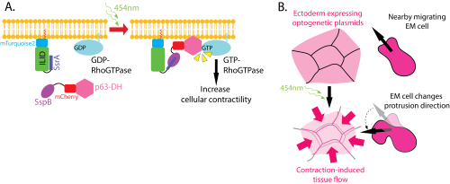
B. How contraction-induced tissue flow biases a nearby immune cell’s protrusion.
One final thought
To me, this project has been the definition of being curiosity-driven. There are many more moments that would make a good nerdy story time over drinks, but I want to give my biggest thanks to my boss, Roberto Mayor, who gave me complete freedom to explore and lead this project. From someone who had many big dreams but was always scared of never getting there (and was absolutely terrified of frogs) to someone who is much more confident to dare to dream bigger, that is how working in Roberto’s lab has changed me. I also want to thank each of my lab mates and all the friends I have made along the way, who have made my life in London much less lonely, being so far away from my family in Glasgow and Vietnam. They truly are the best bunch of people in life for free. And of course, if you know me well, you would know that it wouldn’t be my piece without me thanking Taylor Swift and her amazing music catalogue that pulls me through some of the hardest days! I also hope that I will be able to continue with this line of research into the biomechanics of immune cells within native tissue. Even just a simple video of immune cells migrating through ectodermal cells contains so much biology to be discovered, and I cannot wait to dive deeper into it, either through an organoid approach or another model system like the zebrafish!
And to the younger scientists who are still reading, if I have to give you a few pieces of advice: connect with people in your field, whether it’s online or in person; you never know when you’ll need their expertise; don’t be afraid to take the risk because you never know where it will lead you; learn to adapt, learn to change, let the science guide you; and finally, be like water!
References
1. Costa, R. M. B., Soto, X., Chen, Y., Zorn, A. M. & Amaya, E. spib is required for primitive myeloid development in Xenopus. Blood 112, 2287–2296 (2008).
2. Costa, R., Chen, Y., Paredes, R. & Amaya, E. Labeling Primitive Myeloid Progenitor Cells in Xenopus. in Progenitor Cells: Methods and Protocols (eds Mace, K. A. & Braun, K. M.) 141–155 (Humana Press, Totowa, NJ, 2012). doi:10.1007/978-1-61779-980-8_11.
3. Bera, K. et al. Extracellular fluid viscosity enhances cell migration and cancer dissemination. Nature 611, 365–373 (2022).
4. Mahlandt, E. K. et al. Opto-RhoGEFs, an optimized optogenetic toolbox to reversibly control Rho GTPase activity on a global to subcellular scale, enabling precise control over vascular endothelial barrier strength. Elife 12, RP84364 (2023).
5. Le, H. A., Hartmann, J., Alert, R. & Mayor, R. Tissue flow acts as a guidance cue for immune cell polarization and directional migration. Nat Cell Biol 28, 2008–2026 (2026).
6. Le, A. H. et al. CYRI-A limits invasive migration through macropinosome formation and integrin uptake regulation. Journal of Cell Biology 220, e202012114 (2021).
7. Cruz, M. R., Paixão, T., Coelho, J. & Norden, C. Tissue lamination state switches neuronal translocation from active to passive in the vertebrate retina. Preprint at https://doi.org/10.64898/2026.07.02.736014 (2026).
Get involved
Create an account or log in to post your story on the Node.
Sign up for emails
Subscribe to our mailing lists.