

Flowing into form: quantitative tissue morphogenesis by the sea in Alibaug
Posted by Debasmita Mitra, on 15 November 2026
Written by Debasmita Mitra and Sameer Thukral
Dheere dheere re mana, dheere sab kuch hoye; maali seenche sau ghada, ritu aaye phal hoye.
Slowly, O mind, all things come in their own time; the gardener may pour a hundred pots of water, but the fruit comes only in its season. — Kabir (15th-century Indian poet)
Some places have a way of calling you back. The Fountainhead is a boutique resort in Alibaug, on the coast just south of Mumbai, where the sea is never far away. In 2017, it hosted Cell Biology and Physics of Morphogenesis, a small meeting, organised by the Tata Institute of Fundamental Research, Mumbai, India, with one of us (Sameer) in the audience as a PhD student. Nine years later, from 28 September to 1 October 2026, the same venue hosted the EMBO Workshop Quantitative tissue morphogenesis – models, manipulations and mechanisms (QTIM), organised by the same host institute, Tata Institute of Fundamental Research, Mumbai, India.
The fruit finally had its season.
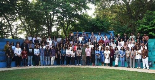
Group photo with all the participants and volunteers of QTIM 2026 at the Fountainhead
A meeting grown at home
The roots of this meeting were apparent in its programme. The small but strong morphogenesis community in India contributed ten of the 32 talks, from TIFR in Mumbai and Hyderabad, NCBS and ICTS in Bengaluru, and the IISERs in Pune, Tirupati and Thiruvananthapuram, alongside speakers from Europe, Japan, Singapore and the US. If the talks gave us a glimpse of the crème de la crème of morphogenesis research in India, the posters extended that richness. Most of the roughly 50 posters came from Indian labs, and their author lists were a pleasure to read: theorists from ICTS and the Simons Centre, engineers from the IITs, developmental biologists from Agharkar Research Institute to InStem.
The meeting ran as a single track, with a welcome change: selected poster presenters gave three-minute flash talks, giving them a stage and a moment to shine. The poster sessions showcased ongoing work from a growing morphogenesis community – from exploring how patterns and shapes arise, using models of Turing patterns, Notch-Delta inhibition, heterochrony and examining how forces and material properties sculpt tissues, to how signalling and cell fate coordinate form and function, in zebrafish epidermis, palate and fins, Drosophila gastrulation, as well as in the developing cochlea, vasculature and heart.
Sensing force, and deciding what to do with it
One thread running through many talks was when and where cells read forces and use them as information, not just as an output of development. Carl-Philipp Heisenberg (ISTA) showed how the zebrafish enveloping layer keeps its tension constant even as it spreads, sensing stretch at its apical surface through Kibra and aPKC, a little like a violinist retuning mid-concert so the pitch stays true. Roberto Mayor’s (UCL) keynote lecture turned to how conversations between neighbouring tissues lead to each building the other: compression during neural plate bending switches on MMP14 in neural crest cells and sets them migrating, and the migrating crest in turn helps the neural tube close.
Tamal Das (TIFR Hyderabad) asked us to look inward, arguing that such decisions are also made in how organelles are organised, which was taken forward by Richa Rikhy (IISER Pune), who showed how membrane recycling drives formation of the first epithelial cells in Drosophila embryos. Sara Wickström’s (MPI for Molecular Biomedicine) keynote and Yanlan Mao’s (UCL) talk carried the theme to cell states and to how tissues cope when stretched or wounded.
Forces and materials make shape
If the first thread was about listening to forces, the second was about the material they act on. Stephan Grill (MPI-CBG) presented the actomyosin cortex as an active matter, while Mahendra Sonawane (TIFR Mumbai) discussed how actin-based apical projections are organised. Vijaykumar Krishnamurthy (ICTS) showed that cytokinesis can emerge from feedback between cell shape and contractility alone, making division less a command than a physical instability that biochemistry tunes. Juan Manuel Gomez and Robert Prevedel (EMBL) used Brillouin microscopy, which reads stiffness with light, to show the folding mesoderm of the Drosophila embryo transiently stiffening alongside changes in microtubule order.
Sundar Naganathan (TIFR Mumbai) asked a question we rarely think of: how tissue flows come to a stop, while Chii Jou Chan (Mechanobiology Institute, Singapore) brought fluid mechanics into the growth of ovarian follicles. Priti Agarwal (NCBS) showed how distinct actomyosin architectures build and run the C. elegans gonad, Raj Ladher (NCBS) discussed how distinct tissues in the inner ear, present in the same milieu, are patterned by the actomyosin cortex, and Sameer Thukral (RIKEN-BDR) presented how the yolk, a phase-separated compartment, mechanically constrains blastoderm formation and gastrulation in flies. Sebastian Streichan showed that most large-scale features can be explained via generalized and coarse-grained physical models by taking examples of morphogenetic events from Drosophila gastrulation to in vitro tissue cultures.
Numbers as a way of seeing
The third thread was omnipresent: measuring biology carefully does not just answer a question; it can change which question you ask. Yoshihiro Morishita (RIKEN BDR) showed that chick and frog limb buds deform in strikingly similar ways once you scale out differences in size and timing; Anna Kicheva (ISTA) combined quantitative assays and models to ask how the neural tube sets up and reads opposing morphogen gradients, while Ina Sonnen demonstrated beautifully how intestinal cell states are encoded in frequencies of transcription factor oscillations. Shigeo Hayashi talked about how careful quantitative analysis of static images of the adult Drosophila cuticle through electron microscopy can reveal how stunning patterns emerge.
Rita Mateus (MPI-CBG) asked how growing fins find their way back to the right size after injury, and Daria Razmadze (University of Helsinki) used Bayesian modelling to compare how lizard teeth acquire their cusps. Martin Weigert (TU Dresden) reminded us that none of this happens without tools such as machine-learning methods for measuring across space and time.
The sea, the food, and the next meeting
Talks are only half of any meeting; the other half happens between them, and here the setting did much of the work. The meeting size was just right for diverse people to meet each other, and the food was excellent, spiced perfectly for all palates. Science, it turns out, travels well on a full stomach.
If this meeting was the flower of the one in 2018, its seeds are already scattered: in the participants who stood up for three minutes of flash talks, in the two evenings of posters, and in all who travelled to Alibaug for the first time. Many of them will meet again to celebrate the 50th Anniversary of the Indian Society of Developmental Biologists (InSDB) at the joint InSDB–ISDB meeting in Goa in 2029, where the same morphogenesis community will be represented in its sessions. If you have never come to a scientific meeting in India, come and see it bloom in 2029.
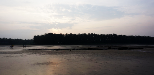
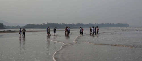
An early morning escape to Awas beach at sunrise provided a fun, refreshing break from the intense scientific sessions.
Afternote: two perspectives
Sameer Thukral (RIKEN BDR, Kobe)
For me, the meeting was a homecoming moment, where I met my PhD mentor Richa Rikhy and labmates, including Debasmita. A conference where you pay attention to every talk and don’t want to skip any session, even talks you have heard before, was a welcome change considering my shortening attention span. From being a member of the audience in 2018 to having the opportunity to present my work, life indeed goes around.
Debasmita Mitra (TIFR, Mumbai)
As a young researcher just beginning to find my feet in the morphogenesis community, QTIM26 was a truly wonderful experience. The meeting was a melting pot of scientists working on various aspects of morphogenesis from across India and around the world. I loved hearing about the latest research in the field and chatting with people whose work I had only read about. I left with new ideas, new friends, and a deeper sense of belonging to this community.
Debasmita Mitra is a postdoc in the lab of Mahendra Sonawane at TIFR, Mumbai, India, working on the interaction between extracellular matrix remodelling and epithelial morphogenesis. Sameer Thukral is a postdoc in the lab of Yu-Chiun Wang at RIKEN BDR, Kobe, Japan, working on the mechanics of yolk–blastoderm interactions.
The observations made here are the authors’ own and do not reflect the opinions of their employers. This article was written in collaboration with Claude AI.
Get involved
Create an account or log in to post your story on the Node.
Sign up for emails
Subscribe to our mailing lists.