Charting new territory: mapping the cell types in the octopus brain
Posted by Ruth Styfhals, on 17 February 2023
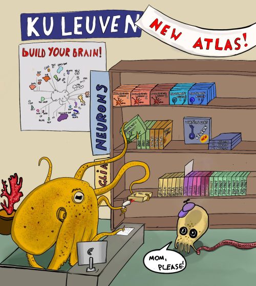
Ruth Styfhals and Dr. Eve Seuntjens at the KU Leuven, Belgium, recently published a cell type atlas of a developing octopus brain in Nature Communications. The team behind the paper was diverse, bringing together the different expertise needed to pull off this challenging project. The authors used both single cell and single nuclei RNA sequencing to identify the different cell types within dissected brains of recently hatched Octopus vulgaris (common octopus). They identified the location of several of these cell types within the brain and compared their molecular profile with brain cell types in other species.
How did you get started on this project?
In general, our lab is interested in understanding the molecular and cellular mechanisms of complex brain development. By making genetically modified mice to model human neurodevelopmental disorders we tried to identify the mechanism behind these disorders. Our focus was on mutations in Protocadherin genes. The latter were known to be important in vertebrates for cell sorting and neuronal wiring. When the first octopus genome was published in 2015, it revealed massive expansions in genes encoding Protocadherins (Albertin et al., 2015). Our lab got intrigued – could these molecules represent an evolutionary convergent mechanism to build and wire up the complex octopus brain as well? We chose to work on Octopus vulgaris, which is easy to obtain in large quantities as eggs. Before being able to study this question, we first needed to set up a system to keep and hatch the Octopus eggs in the lab (Deryckere, Styfhals, Vidal, Almansa, & Seuntjens, 2020), and we described brain development using modern technologies such as light sheet imaging (Deryckere, Styfhals, Elagoz, Maes, & Seuntjens, 2021). After identifying the neurogenic niche during embryonic development, one very important missing piece of information was molecular knowledge about cell type diversity in the brain. What was the end point of embryonic brain development, and how many cell types were present in this alien brain? Our angle was a developmental one, and we therefore focused on the brain of freshly hatched ‘paralarvae’, which is the swimming intermediate stage that grows over the course of about 5 weeks into a juvenile that settles and adopts the benthic lifestyle of adult Octopus vulgaris.

What was already known about the cell types in the octopus brain?
In 1971, a detailed overview of the anatomy of the adult nervous system of Octopus vulgaris was published (Young, 1971). Therefore, we had a good idea of the different brain lobes, their connections and their function in the adult. In addition, nuclear sizes and the morphology of different cell types were described. Nothing was really known about the number of cell types, what molecular markers these cell types had and how to link molecular types to “morphotypes” present in the brain. Right after hatching, the brain only has about 200,000 cells – therefore it still needs to multiply 1000-fold to reach the cell numbers present in the adult nervous system, which is around two hundred million. Therefore, we were not sure whether we could really compare this hatchling brain with the adult one. Molecular knowledge at the embryonic or larval stage was very limited to studies on selected transcription factor gene expression, often not really at cellular resolution. We also knew that certain neurotransmitters should be present, based on the adult work. We could not even guess how many clusters to expect, and did not have any marker genes to annotate clusters.
Can you summarize your findings?
Our results revealed that the octopus hatchling brain already contains a stunning diversity of cell types. These cell types are often organized according to molecular profile to appear in specific locations showing that this brain is already highly organized. We found that most of the cells are neurons, but there are also distinct glial cell types, and some seem to be spatially confined. We tried to distinguish ancestral cell types from novel cell types by using comparisons to mouse and Drosophila brains, and found that cells of octopus vertical lobe (the brain structure necessary for memory and learning) are transcriptionally similar to cell of the fly mushroom body, indicating functional convergence. We also found that novel Octopus-specific genes, like Protocadherins, are used to delineate specific cell types that might represent evolutionary novel cell types. Working with an unusual species brought additional challenges. A first key step was getting sufficient high-quality samples, by performing micro-dissection, optimizing isolation of cells and having expert help with nuclei isolation. A second key step was to ameliorate, in a significant manner, the gene model annotation of the genome, even when this genome already had a chromosome-level assembly. Using long-read Iso-seq and FLAM-seq data, we could extend 3’ ends in a data-driven manner, increase mapping statistics and more than double the amount of data. A third important step was the spatial mapping using hybridization chain reaction, a very powerful method for revealing gene expression in situ in non-model species. This enabled us to create an initial map of the cellular diversity.
When doing the research, did you have any particular result or eureka moment that has stuck with you?
When comparing octopus brain cell types to mouse and fly brain cell types, we initially didn’t really expect to find anything useful, because of the immense evolutionary distance (the ancestor of octopus and mouse lived about 600 million years ago). It was striking to see that glial cells in all three species were alike, as were neuronal cells important for memory and learning in fly and octopus. This was most amazing, to see evolutionary conservation -or convergence- on a cellular level!
And what about the flipside: any moments of frustration or despair?
Starting up an entirely new non-model, marine aquatic animal culture in a lab with background mainly in mouse development was challenging and took its time. Many grant reviewers were not convinced we were able to pull this off, leading to most grants being rejected. This meant we needed to be very creative with our minimal resources, and we were dependent on help from more fortunate collaborators who did see the innovation and the potential of the idea. Firstly, Stein Aerts, who co-founded FlyCellAtlas, chipped in some of his resources to perform a bold dual single-cell and single-nuclei experiment. Stein is a long-time collaborator and his no-nonsense attitude kept us focused on the goal: to get an initial octopus brain cell atlas. Secondly, Nikolaus Rajewsky developed an interest into octopus brain RNA profiles, and attracted the hyper-dedicated and talented master student Grygoriy Zolotarov to work on this project. We teamed up and were able to massively ameliorate annotation and gene models which more than doubled the amount of usable data. Thirdly, we did not start from the void. Previous collaborations with Gregory Maes, at that time IOF manager at the genomics core facility of KU Leuven, had yielded isoseq long read transcriptome data. Last but not least, our long-standing collaborator Eduardo Almansa made sure we had access to egg clutches and provided them to us at no charge. We wanted to give a shout out to these key people and their generosity; without them this story would not have existed.
What is next for you/the lab after this paper?
Ruth (first author) is finishing her PhD and is currently looking forward to working on neural development in even more unknown, weirder organisms, which have a less complex brain than that of the octopus.
Where will this story take the lab?
This project for sure has opened up a number of future research lines. Having a molecular view on cell types, the next challenge is to link these types to the morphotypes found by JZ Young and others. Another challenge is to understand how this diversity is generated during development: is there a spatial and temporal logic to these cell types? Do neurons and glia have a common stem cell or not? What transcription factors and signaling molecules determine cell fate and migration? How does this brain grow beyond hatching? Are larval cell types retained or replaced? How are these cell types wired up? And how do they lead to (innate) behaviors one can observe in the paralarval phase? There are still many unknowns, but with this molecular profiling of cell types, we can now better formulate hypotheses that might bring new insights into the function of this enigmatic big brain.
by Ruth Styfhals and Dr. Eve Seuntjens (eve.seuntjens@kuleuven.be)
References
Albertin, C. B., Simakov, O., Mitros, T., Yan Wang, Z., Pungor, J. R., Edsinger-gonzales, E., … Rokhsar, D. S. (2015). The octopus genome and the evolution of cephalopod neural and morphological novelties. Nature, 524, 220–225. https://doi.org/10.1038/nature14668
Deryckere, A., Styfhals, R., Elagoz, A. M., Maes, G. E., & Seuntjens, E. (2021). Identification of neural progenitor cells and their progeny reveals long distance migration in the developing octopus brain. ELife, 1–32. Retrieved from https://doi.org/10.1101/2021.03.29.437526
Deryckere, A., Styfhals, R., Vidal, E. A. G., Almansa, E., & Seuntjens, E. (2020). A practical staging atlas to study embryonic development of Octopus vulgaris under controlled laboratory conditions. BMC Developmental Biology, 20(6), 1–18. https://doi.org/10.1101/2020.01.13.903922
Styfhals, R., Zolotarov, G., Hulselmans, G., Spanier, K. I., Poovathingal, S., Elagoz, A. M., … Seuntjens, E. (2022). Cell type diversity in a developing octopus brain. Nature Communications, 13(7392), 1–17. https://doi.org/10.1038/s41467-022-35198-1
Young, J. Z. (1971). The anatomy of the nervous system of Octopus vulgaris. London, UK: Oxford University Press.


 (No Ratings Yet)