In Development this week (Vol. 143, Issue 22)
Posted by the Node, on 15 November 2016
Here are the highlights from the current issue of Development:
A postnatal model for Zika virus infection
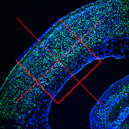
Zika virus (ZIKV) infection results in  embryonic microcephaly and has been declared a global health emergency by the World Health Organization. Disruption of the neural progenitor cells is considered to be the major cause of microcephaly; however, the fate of other cell types, including differentiated neurons and vascular cells, remains unknown. Since infected mouse embryos die perinatally, it also remains unknown whether ZIKV infection can cause postnatal microcephaly in animal models. Now, on p. 4127, Jian-Fu Chen and colleagues report a postnatal model for ZIKV infection using intracerebral inoculation of embryonic brains with the ZIKV. The infected pups survive after birth and show postnatal microcephaly, which bears relevance for a better understanding of the microcephaly observed in ZIKV-infected newborn humans. In addition to microcephaly, the postnatal mouse model recapitulates several aspects of fetal brain abnormalities associated with ZIKV in humans, including extensive neuronal apoptosis and loss, axonal rarefaction, corpus callosum diminishment, and reactive astrocyte and microglial cell accumulation. Furthermore, the authors show that ZIKV infection leads to increased vessel density and vessel diameter, and causes blood–brain barrier leakage in the developing brain. While further research is required to better characterise the approach, the development of a postnatal model for ZIKV infection is an important step forward in understanding this disease, and the findings reported by the authors offer novel insight into the pathology of ZIKV infection in the postnatal setting.
New network for tooth development: Sox2 bites back
 Initiation and subsequent growth of the mammalian tooth depends on distinct populations of epithelial and mesenchymal stem cells located in the labial cervical loop (LaCL) and the neurovascular bundle, respectively. In rodents, Sox2 marks the dental epithelial stem cells (DESCs) and has been shown to be an important regulator of tooth development, but the molecular mechanism by which this occurs has not been determined. In this issue (p. 4115), Brad Amendt and colleagues uncover a Pitx2/Sox2/Lef-1 network that controls the epithelial stem cell niche in the continuously erupting rodent incisor. The authors demonstrate that Sox2 is necessary for the maintenance of the stem cell niche, as inactivation of Sox2 leads to lower incisor arrest, as well as abnormalities in the upper incisor and molar teeth. Conditional overexpression of Lef-1 can partially rescue the Sox2-related defect in incisor growth, possibly owing to increased cell proliferation at embryonic stages and the formation of a new compartment of stem cells in the LaCL. The authors also provide evidence for physical interaction between Pitx2 and Sox2, and show how both factors are core components of the Pitx2/Sox2/Lef-1 network. Together, these findings represent a significant milestone in our understanding of the transcriptional control that defines dental stem cell development and differentiation.
An interview with Kathryn Anderson
 Kathryn Anderson is Professor and Chair of the Developmental Biology Program at the Sloan Kettering Institute in New York. Her lab investigates the genetic networks underlying the patterning and morphogenesis of the early mouse embryo. We caught up with Kathryn at the 2016 Society for Developmental Biology – International Society of Differentiation joint meeting in Boston, where she was awarded the Edwin G. Conklin medal.
Transposable elements in development
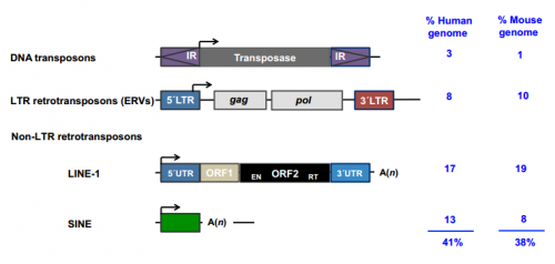
 This Review discusses how and when transposable elements are expressed during development and how they modulate genome architecture, gene regulatory networks and protein function during embryogenesis.
Optimising and improving DamID
Joachim Wittbrodt and colleagues present critical improvements to the DamID protocol improve specificity and sensitivity in determining genome-wide protein-DNA interactions in transient or stable transgenic animal lines.

Genetically encoded, inducible cell death
Reinhard Köster and colleagues present Tamoxifen-induced Caspase activation in zebrafish. This enables fast, efficient and specific cell ablation via targeted apoptosis.

Quantitative stem cell biology: the threat and the glory
This meeting report from Steven Pollard highlights the major advances and emerging trends in quantitative stem cell biology as presented at the 5th annual Cambridge Stem Cell Symposium this year.


 (No Ratings Yet)