Developmental Biology in the Ocean – a crash course in comparative invertebrate embryology
Posted by Nat Clarke, on 8 July 2022
In June 2022, I had the pleasure of teaching a short course on comparative embryology with Chris Lowe and Laurent Formery at Stanford University’s Hopkins Marine Station. Our mission: to take a mix of grad students and postdocs from disciplines across the biosciences, introduce them to diverse developmental mechanisms in a broad sampling of organisms spanning the animal tree of life, and then release them to pursue research projects of their own design.
What is the value of a course like this? And why are marine stations an ideal setting? Simply put, animal diversity showcases countless natural experiments – evolutionary experiments in diverse body plans, novel cell types, complex life history strategies, and more. Since life evolved in the ocean, the seaside environment allows unrivaled exploration of this biodiversity: where else offers ready access to embryos of 15 animal phyla within steps of the classroom?
I could, of course, describe at length what students might get out of such an experience, but instead I’ll give it to you directly from the source. Below, a few of our students, and my co-instructor, share their reflections and favorite moments from the course.
Student Perspectives:
Charlotte Brannon:
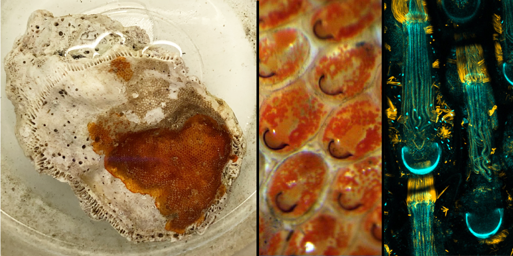
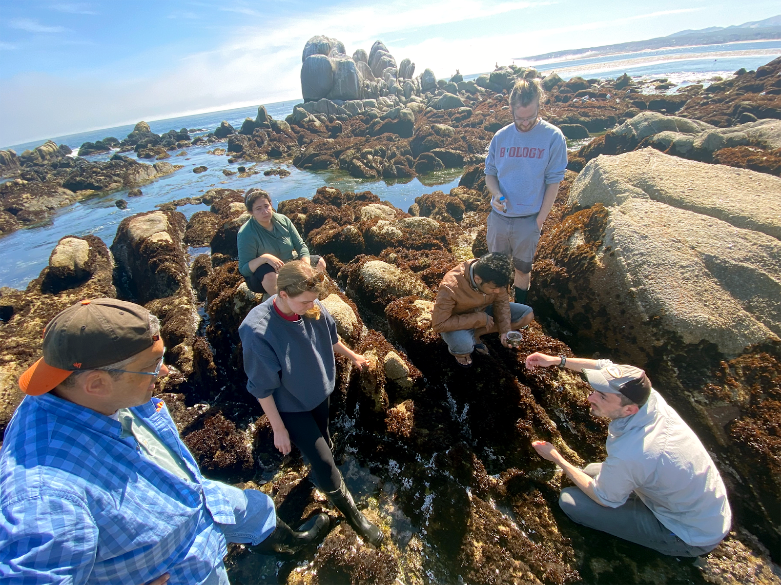
There is so much biology hiding in non-model organisms, especially in the ocean, and it was enlightening to immerse myself in it during this course. Partly, this happened through tidepooling and digging for worms in mud flats. Tidepooling was surprisingly challenging at first. Our instructors could pick up a rock, identify five species on it, and tell you anything you wanted to know about any of them. Meanwhile, the most exciting thing I could find was a floating, white creature which turned out to be… a seagull feather. I ended up collecting a seemingly boring shell with some red stuff encrusted on it. Surprisingly, a quick look under the dissection scope revealed this crusty red stuff to be a Bryozoan colony! Unknowingly, I had collected a fascinating marine invertebrate. Similarly, when we later visited mud flats, a classmate and I found what seemed to us like a very average worm. We later learned that it was actually Leptosynapta albicans – the burrowing sea cucumber. I learned my lesson: when immersed in nature, you must actively try not to find something interesting.
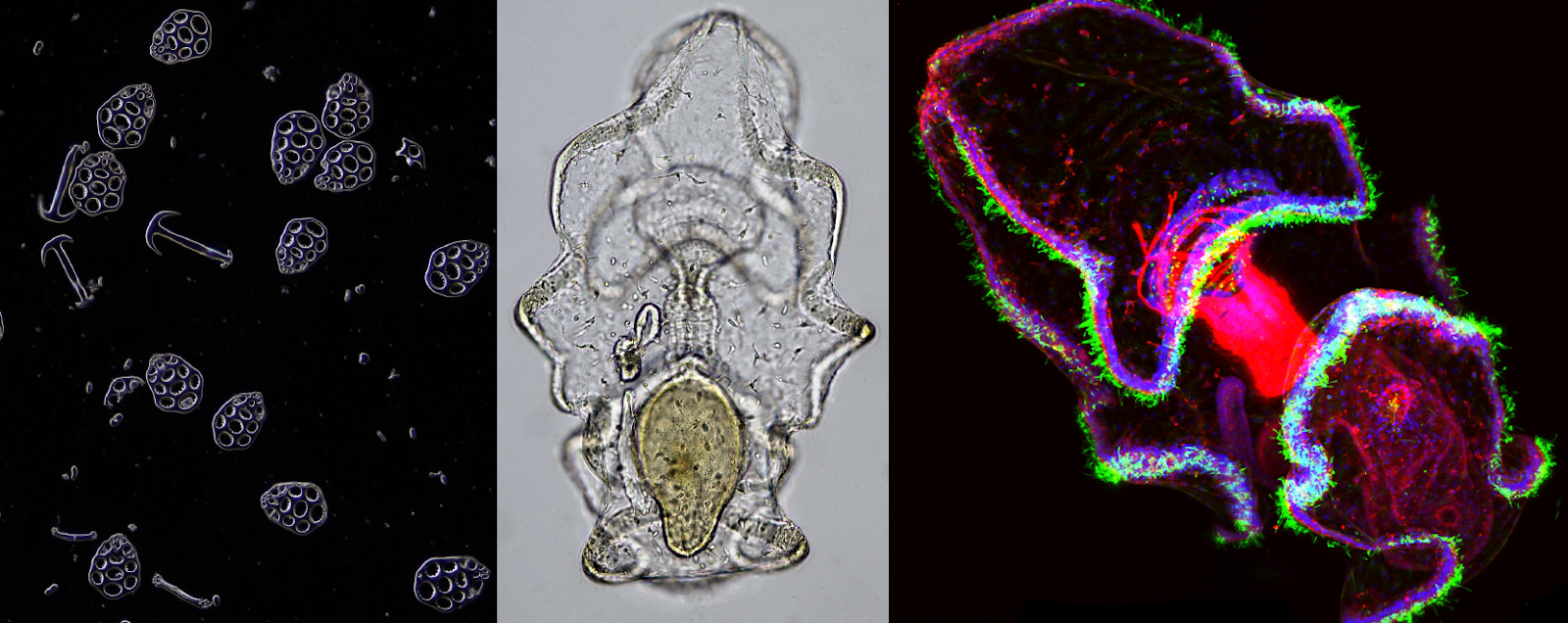
Exposure to a wide range of marine invertebrates made me more excited about developmental biology because it highlighted how much we don’t know! I am excited about all that non-model organisms can teach us, and eager to explore the mechanics of development in a range of systems. Of course, this is easier said than done. As we learned, having the right tools to work with an organism is crucial, and sometimes that means building your own. The short timeline of the course forced us to be creative with our resources and think carefully about the appropriate tools. I also appreciated seeing multiple approaches to the same experiment. For example, our guest instructor, Brady Weissbourd, demonstrated his strategy for injecting Clytia eggs, which differed in many subtle ways from our instructors’ approach for injecting echinoderm eggs. This made me realize how important it is to tailor your experimental approaches to your organism.
Lauren Lubeck:
As a marine research station, Hopkins Marine Station is one of only a few special places where scientists can collect, observe, and perform experiments on a wide diversity of marine invertebrates. It was an incredible experience to be immersed in the marine environment at Hopkins. Unsurprisingly, almost every guest instructor wanted to spend time outside looking for their favorite organisms, and the students went tidepooling on our own many times. We learned the importance of understanding the ecology of our target species. Want choanoflagellates? Look for small, dirty looking pools. Want acoels? Turn over rocks in the sandy sections of tidepools. Want fat innkeeper worms? Look for the holes marking the entrance to their U-shaped burrows. Working where our favorite intertidal invertebrates live created a unique opportunity to learn more about them.

A highlight for me was the connections I made with fellow students. Each student arrived with specific interests and biological questions in mind. I loved that we all found ways to investigate our favorite questions while using species that were new to us. While each of us pursued our original question, we also were spontaneously inspired by a new animal or phenomenon we encountered. For example, Nabor Vazquez Martinez usually studies aspects of the nervous system in C. elegans, but he found Brady Weissbourd’s Clytia jellyfish fascinating and decided to examine their nervous system too. Azalia Martinez Jaimes is interested in stem cell differentiation and found an interesting model in the tunicate Botryllus, which continuously builds new adults from growing buds filled with stem cells.
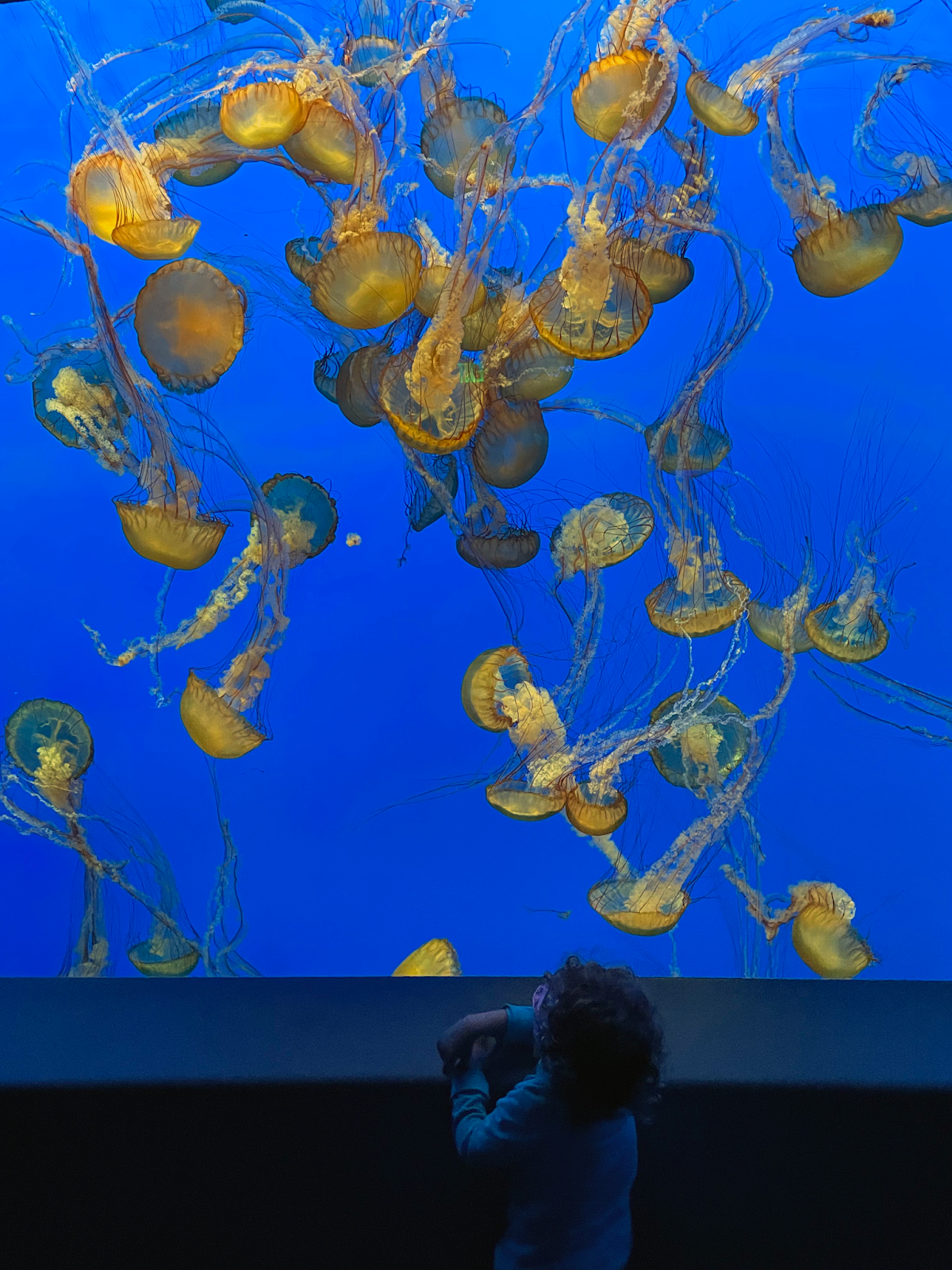
The “Behind the Pipes” tour of the Monterey Bay Aquarium was another highlight. As we were led through the ctenophore facility by Senior Aquarist Wyatt Patry, I learned that they grow the same algae we were feeding our larvae in class, but I was amazed by the massive scale of production. Once we entered the main exhibit to see the ctenophores and cnidarians on display, I was overcome with a combination of awe and excitement. It was moving to see how our research interests meshed with the education and outreach of the Monterey Bay Aquarium.
A. S. Jijumon:
“In all things of nature there is something of the marvelous” — This is a quote from Aristotle that I saw at the Monterey Bay Aquarium during our class visit. In this course, I realized that there is much to learn and explore in marine organisms, and that it can be more straightforward to make novel and important observations in unexplored areas.
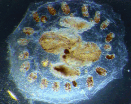
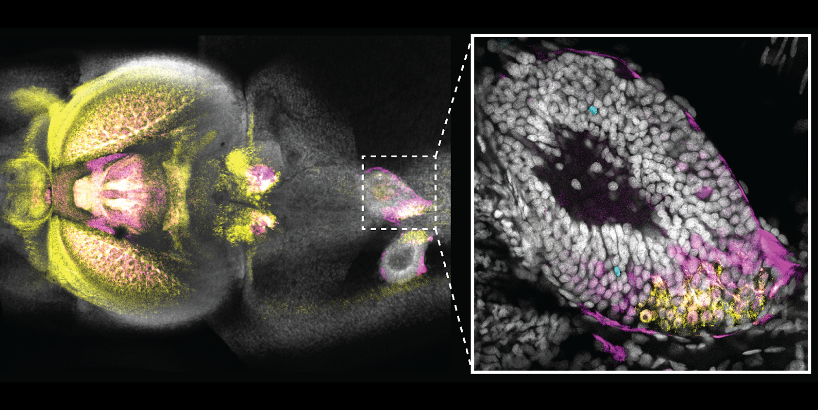
I did my undergraduate education in India, and what I studied in my developmental biology classes were mostly theories and text book images. Through this course, for the first time, I got a practical demonstration of the developmental stages of multiple marine invertebrates and observed their morphogenesis over time. I got a chance to really experience the origin of classical experiments in embryology and developmental biology. Observing the wide diversity of organisms we could collect straight from tidepools, and then performing wet lab experiments on these creatures using microscopy and molecular tools was a fantastic experience.
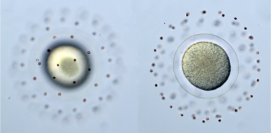
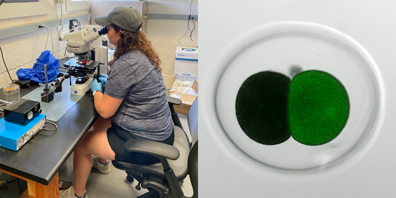
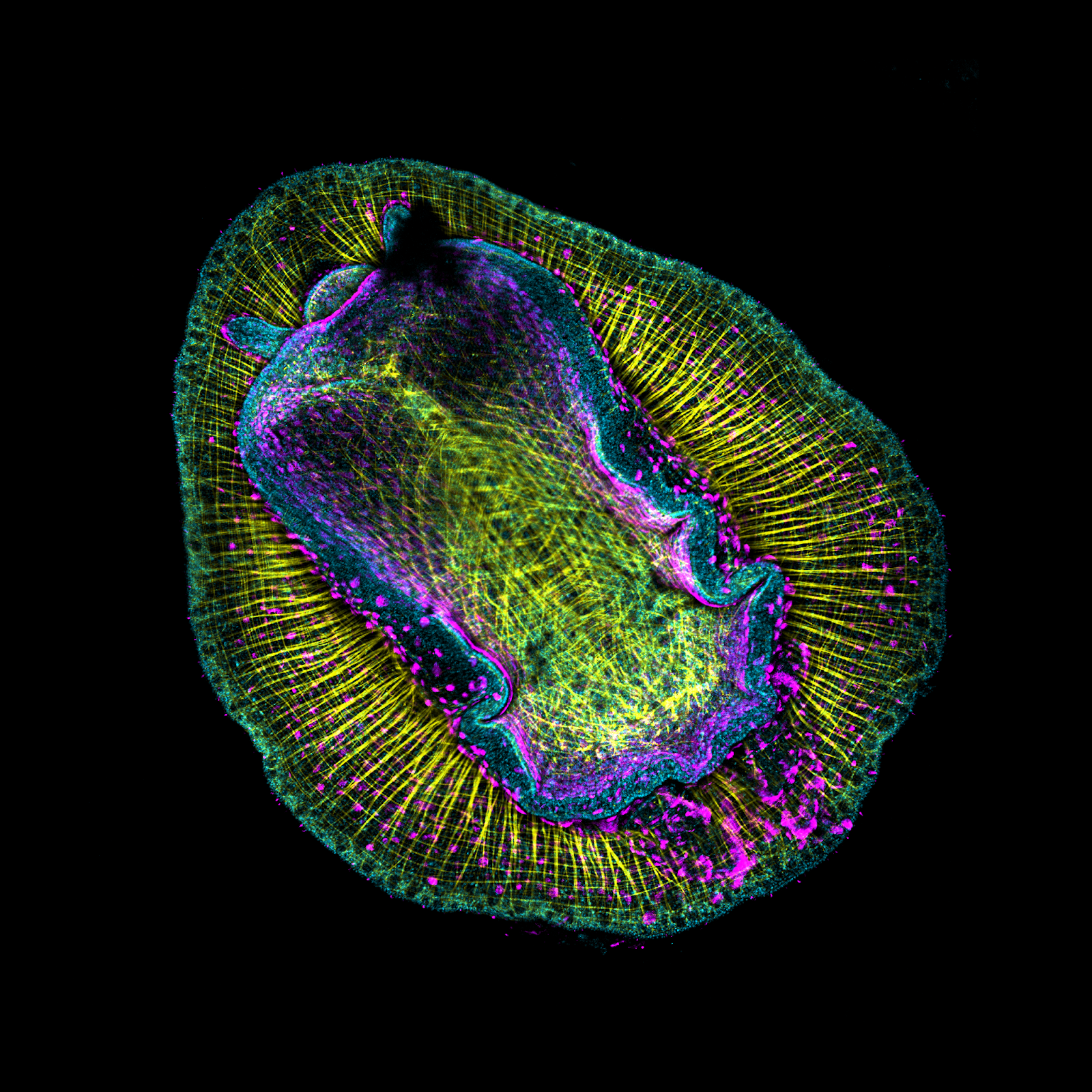
My favorite observation was with the sand dollars (Dendraster excentricus) we worked with on our first day. We collected eggs and sperm by injecting KCl into their gonads. Subsequently, I added diluted sperm onto an egg and watched on the microscope. That was the first time I witnessed the event of fertilization occur in real life, and I felt goosebumps and got the impression that I acquired the power to manipulate life, which was a memorable moment. I would like to share this experience with other science enthusiasts in the future.
Instructor perspective – Laurent Formery:
This course was one of my first teaching experiences, and it was an awesome one. We wanted to promote exploration and experimentation using the incredible resource that we had right outside the classroom – the ocean. From tidepools, mudflats and plankton nets we collected species spanning over a dozen animal phyla (and some of our closest unicellular relatives, too), and we spawned, observed and manipulated them in the classroom. The combinations of the students’ unique skill sets and the array of animals that we collected generated a profusion of discovery – applying well-developed techniques to new questions in new species. During this process I personally learned much more than what I could teach to the students, making this course an enriching experience for me as well. The main message of the course we tried to emphasize was the astonishing diversity of biochemical processes, developmental mechanisms, and ecological strategies waiting to be discovered and documented right there in the ocean. This underscores the importance of protecting the endangered biodiversity of our coastlines, but also the value of supporting basic exploratory research outside the handful of classical biological model systems. One of my favorite examples highlighting the importance of exploring non-model systems was brilliantly told by Dan Rokhsar during his genomics lecture to the class. The recent chromosome mapping of non-model species such as the scallop Patinopecten yessoensis and the jellyfish Rhopilema esculentum enabled the discovery of a fundamental feature of animal evolution: the arrangement of genes on chromosomes (referred to as synteny) is highly conserved and can be traced back to the roots of metazoans (Simakov et al., 2022). The few exceptions to that rule, in which macrosynteny has undergone independent and major reorganization events, are curiously distributed among the metazoan tree: they include the fruit fly Drosophila melanogaster, the nematode Caenorhabditis elegans, and the entire vertebrate clade.
Acknowledgements: A course like this simply cannot run without the support of a team of deeply invested staff and instructors. We want to give a special “thank you!,” to the staff at Hopkins, and to all of our guest instructors – Dan Rokhsar, David Booth, Flora Rutaganira, Christina Zakas, Bo Wang, Brady Weissbourd, Ryan York, Wyatt Patry, Deidre Lyons, Jessica Goodheart, Dominique Bergmann – for making this year’s course a success. We also thank the departments of Biology, Developmental Biology, and BioEngineering, at Stanford for financial support, and Molecular Instruments and Luxendo for generously providing equipment and reagents for the course.
 (4 votes)
Get involved
Create an account or log in to post your story on the Node.
Sign up for emails
Subscribe to our mailing lists.