Euro-BioImaging – Europe’s gateway to biological and biomedical imaging excellence
Posted by Marianna Poli, on 4 September 2023
Do you feel as if a novel imaging approach would give you new insights into your sample or provide a new way to answer your research questions? Imagine you could take your research question to any institute and work with the experts to unlock the power of imaging technologies and get new data and insights. Well, you can – with Euro-BioImaging.
What is Euro-BioImaging?
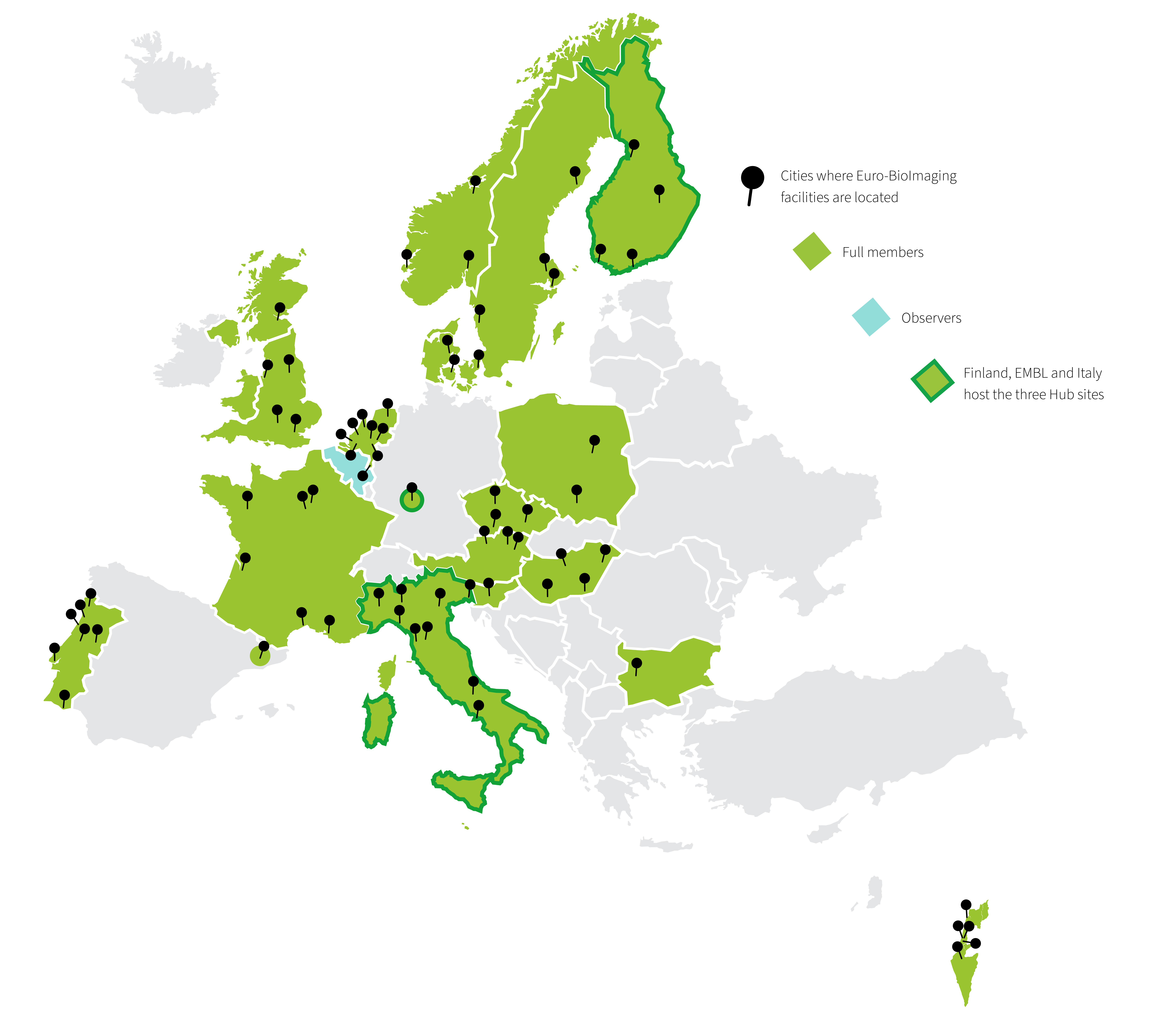
Euro-BioImaging is the European research infrastructure for biological and biomedical imaging that functions as the gateway to over 170 world-class imaging facilities across Europe. Through Euro-BioImaging any researcher, from anywhere around the world, can get access to imaging technologies, image data services and training.
It builds on a set of already existing national and international facilities of excellence in imaging technologies, the Euro-BioImaging Nodes, which provide physical or remote access to imaging technologies, deliver training and support the users at all the stages of their research projects.
Together, over 500 core facility staff work at Euro-BioImaging Nodes and support researchers. We are a fabulous resource for researchers across scale, from atoms to humans, and in diverse disciplines in the life sciences.
How does Euro-BioImaging support researchers?
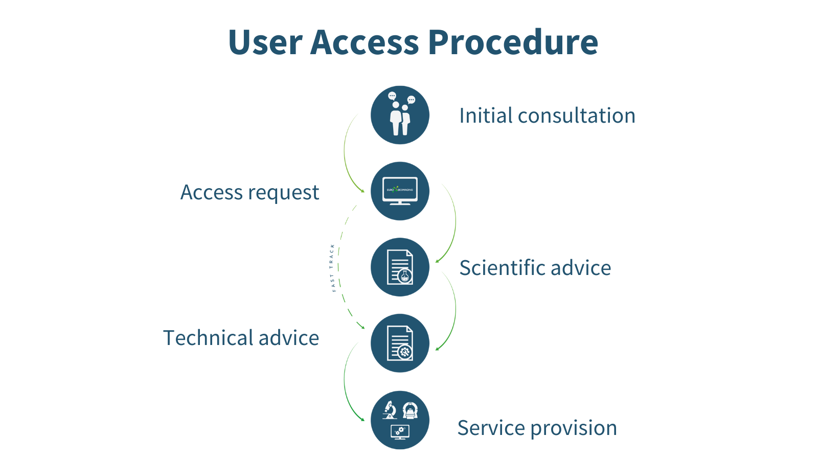
Every researcher, independent of research area, level of expertise, and geographical location, can apply for Euro-BioImaging services whenever they have a project requiring imaging technologies or expertise which they do not have ready access to at their home institute. The expert staff at the Euro-BioImaging Hub can help future users choose the right technology and facility for their research question in the first step of the User Access procedure. Applying for user access is a highly collaborative process in which a researcher has multiple opportunities to hone their experiment and get scientific and technical input from reviewers and technical experts at the imaging facilities before the application is accepted and service provision begins.
An accepted Euro-BioImaging project can be a game-changer. It democratizes access to high-end imaging technologies to push a research question and publication to the next level, and is a starting block towards acquiring new skills, expertise and scientific insight. Euro-BioImaging Users benefit from the expertise of the imaging scientists at the Euro-BioImaging facilities when it comes to sample preparation, experimental set-up and data analysis. Depending on the scope of the project and selected technology, users may also learn skills that they can take back to their home institute.
These skills open doors, especially for early career researchers. Being selected for Euro-BioImaging user access is also a good endorsement of the underlying scientific question or application. Undertaking a project at a Euro-BioImaging facility proves a researcher’s ability to plan and carry out an experiment from start to end.
What imaging technologies are available?
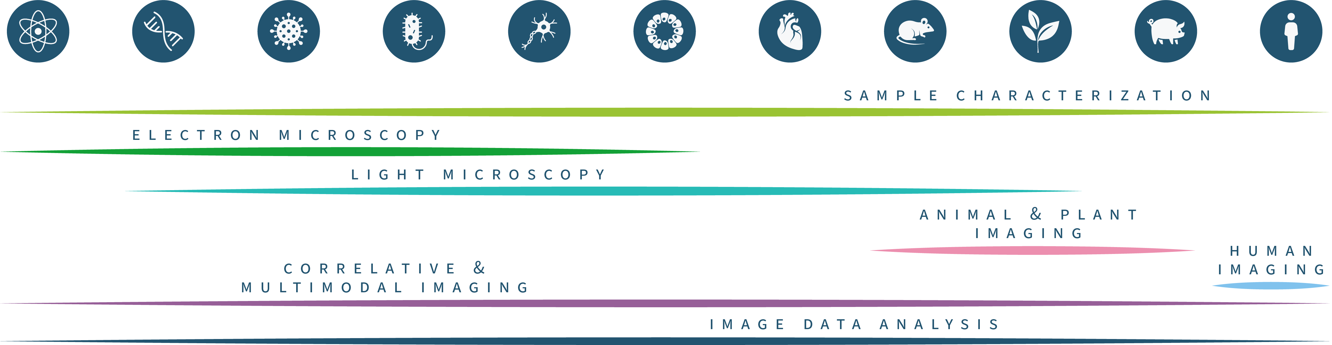
Through the large number of facilities, Euro-BioImaging can offer access to the full range of imaging technologies in the biological and biomedical imaging field. Our technology portfolio covers everything from the nano- to the tissue- and organism scale. We are constantly adding new technologies, making sure that the latest cutting-edge imaging technologies, such as MINFLUX and spatial transcriptomics, are available in open access to all researchers.
Harnessing the imaging revolution
The Euro-Bioimaging technology portfolio ranges from light and electron microscopy on the biological imaging side to an expanding range of applications of biomedical imaging, from plant and ex-vivo imaging to animal and human imaging applications.
Electron Microscopy
Our Electron Microscopy portfolio covers cryo-EM techniques for ultrastructural exploration, such as cryo-electron tomography (cryo-ET), as well as the full complement of volume EM techniques, such as FIB-SEM, Array Tomography and Serial Blockface SEM. Many of our facilities also specialise in correlative methods, Correlative X-ray Imaging and EM (CXEM) and correlative light and electron microscopy (CLEM).
Light Microscopy
In light microscopy, our Nodes offer everything from basic confocal microscopy up to single molecule location approaches and intravital imaging. Our light microscopy techniques allow for 3D live cell imaging, tracking, high content screening, and include a variety of functional imaging techniques to explore protein dynamics in living cells. Recently we have added a number of new and highly requested methods, such as MINFLUX, Single Particle tracking and Lattice Lightsheet microscopy to our portfolio.
Model Systems
Euro-BioImaging facilities also offer access and support with a wide range of model systems and how to get the best imaging results out of them, from Drosophila and zebrafish to mouse embryos and organoid systems. Here access to instruments is complemented by technical expertise of facility staff, to support specialized sample handling.
Support Technologies
And of course, we also provide access to adaptive and support technologies, such as laser- based microdissection, Feedback Microscopy, high-speed imaging, microscopes at high biosafety levels, and specialized sample preparation methods, such as Tissue Clearing and Expansion Microscopy.
Euro-BioImaging can also support you if you want to explore the physical and chemical properties of your samples, through access to a range of methods such as MassSpec Imaging, Atomic Force Microscopy, and chemical imaging, such as µ-XRF and μ-PIXE.
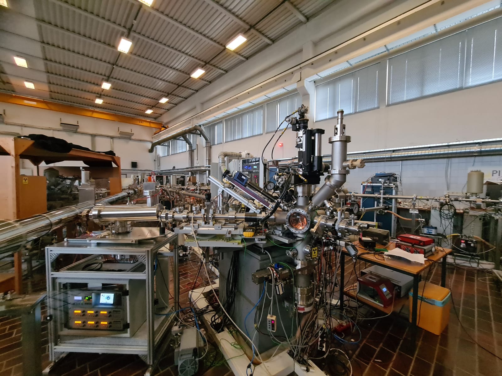
Figure 4: Micro-PIXE at the Jožef Stefan Institute, part of our SiMBION Node in Slovenia
How will Euro-BioImaging enhance my research?
So, when you read about a cool, new microscopy method in the literature, you can now allow yourself, not just to imagine, but test the impact that method could have on your research question. If it’s a technology that Euro-BioImaging offers, you can always apply. Because the idea behind Euro-BioImaging is to make the best imaging resources available to all researchers, providing new answers to scientific questions and increasing the impact of research.
What about image data analysis?
Image data analysis is an integral part of any experiment and is therefore usually integrated into the experimental concept at Euro-BioImaging at an early stage. Experts at the Nodes help users extract their data and set up image analysis pipelines, typically preparing for image analysis and data extraction, sometimes even before the actual experiment begins. In addition, Euro-BioImaging offers its users Image Data Analysis (IDA) as a stand-alone service through expert Image Analysts at the Nodes, irrespective of where the image data was acquired.
Users can contact our Nodes when they need:
• Biological and biomedical image data analysis support
• Image registration, segmentation, tracking and more
• Data workflows, bespoke analysis tools and machine learning methods
• Access to high performance computing and specialized software
How can I apply?
You can apply to carry out a project at a Euro-BioImaging Node via our website. Our website provides instructions on how to access here: https://www.eurobioimaging.eu/about-us/how-to-access
Before you submit a proposal, feel free to browse our technologies and services, view our user stories to get a feel for what Euro-BioImaging can offer, and reach out to us for help in choosing the right imaging method.
What other opportunities are available?
Training
If you’re not ready to undertake a full experiment with Euro-BioImaging, why not start with a training course? Euro-BioImaging Nodes offer a wide range of training courses – covering the full spectrum of biological and biomedical imaging technologies as well as sample preparation and handling, and image data analysis. Some courses are taught remotely and virtually, increasing their accessibility. Taking a course at a Euro-BioImaging facility is a great way to learn a new skill or improve your technique and to build your network. Here’s an overview of training courses available at Euro-Bioimaging Nodes.
Community building
In addition, Euro-BioImaging organizes regular events focusing on imaging for the benefit of the entire community. You are welcome to join our weekly “Virtual Pub” – a free weekly lecture series, open to all imaging enthusiasts. Topics include new biological and biomedical imaging technologies, image analysis, and other topics of interest.
In addition, we organize a “User Forum” twice a year to highlight the importance of imaging to different research areas. These events feature keynote presentations from prominent scientists as well as presentations from users at our Nodes. We usually record them and make the content available on our YouTube channel.
And finally, we are present at many conferences and community events. So we hope to meet with you and talk face-to-face about the wonderful opportunities Euro-BioImaging can provide. Until we meet in person, you can always reach out to us by email info@eurobiomaging.eu or sign up for our Newsletter to stay informed.
 (2 votes)
Get involved
Create an account or log in to post your story on the Node.
Sign up for emails
Subscribe to our mailing lists.