Featured image with Julia Peloggia de Castro: the Node–FocalPlane image competition
Posted by the Node, on 3 April 2025
To accompany the Biologists @ 100 conference, we’ve partnered with FocalPlane to bring to you an image competition. We shortlisted 15 images and asked you to vote for your favourite online and in person at the conference. On the final day of the conference, we announced the top3 images of the competition.
In this ‘Featured image’ post, we find out more about the story behind Julia Peloggia de Castro’s image, which was one of the runners-up in the competition.

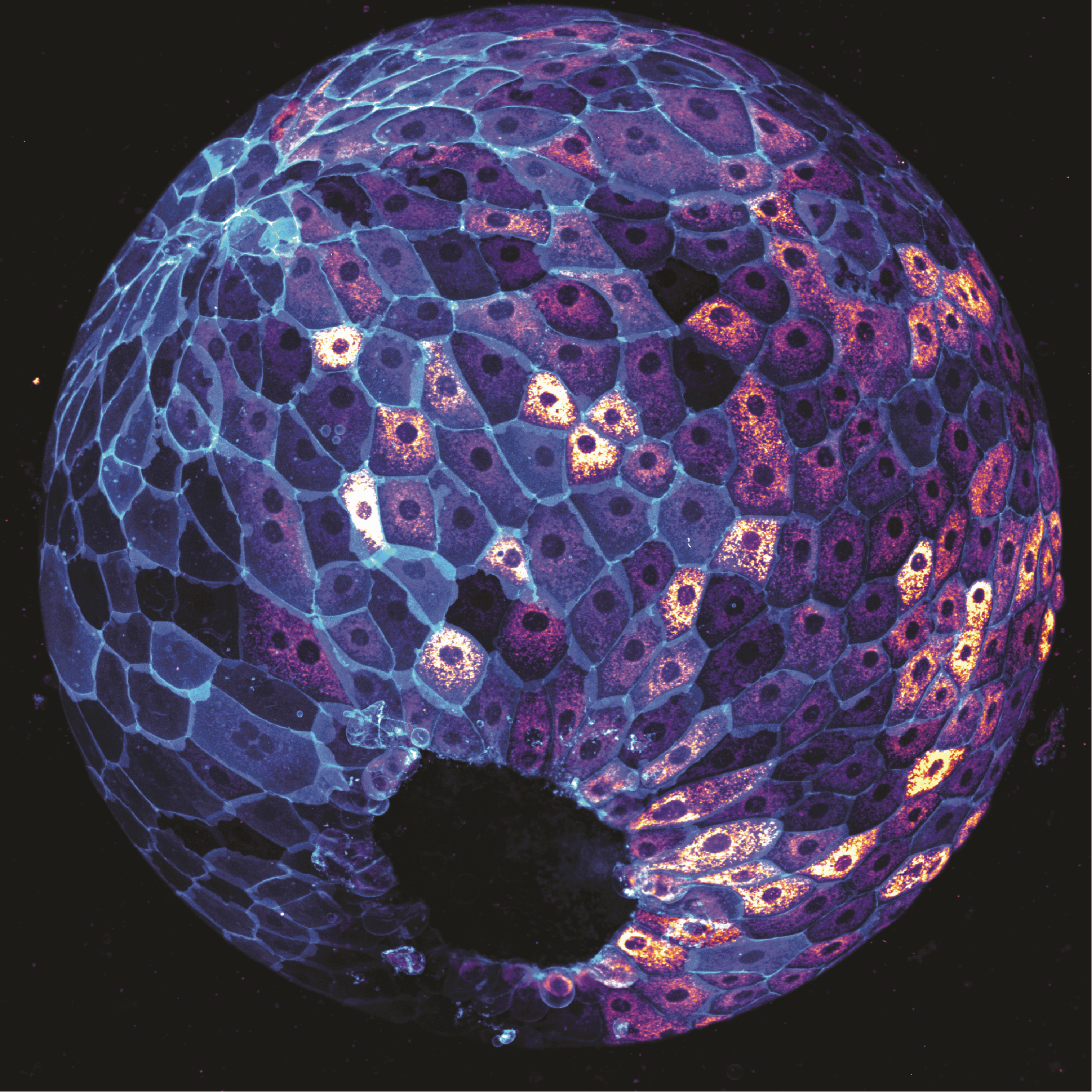
The image depicts a zebrafish embryo at 9 hours post-fertilisation on a lateral view. Cells are stained with MitoTracker, which labels active mitochondria, and cell membranes are labelled in cyan with a EGFP transgenic membrane tag. Image was taken using a 20x objective on a spinning disk confocal microscope.
What is your background?
I got my bachelor’s degree at the University of São Paulo, in Brazil. I majored in Biological Sciences with a focus on genetics and molecular biology. I then pursued my PhD at the Stowers Institute for Medical Research in Kansas City (USA), where I studied zebrafish development and phenotypic plasticity under the mentorship of Dr. Tatjana Piotrowski. I am now a postdoctoral fellow at University of California, Los Angeles (USA) working with Dr. Heather Christofk.
What are you currently working on?
My current interests are in developmental metabolism, and how maternal metabolic states impact mouse embryonic development.
Can you tell us more about the story behind the image that you submitted to the image competition?
This image shows a zebrafish embryo after a few hours of development. One of my favorite things about zebrafish is how fast they develop, giving us the opportunity to watch it —and image it— in real time. This particular image was taken late at night in the laboratory while I was testing a reagent and optimizing imaging parameters before running a larger experiment later that week. The result was definitely worth staying late for!
What is your favourite technique?
I really appreciate any type of microscopy. From the foldscopes to confocal and electron microscopes. Looking at cells never gets old!
What excites you the most in the field of developmental and stem cell biology?
I think this is a very exciting moment in developmental biology research. The last decade has brought many technical revolutions and more fields are bridging towards developmental biology, generating new questions and new ways to approach them. This makes me think we are poised to discover new biological principles soon.


 (No Ratings Yet)