The backbone of stem cell derived embryos — featured image from the Node-BSDB virtual art exhibition
Posted by the Node, on 20 December 2023
In the recent BSDB-the Node virtual art exhibition, Christoph Markus Haefelfinger’s ‘The backbone of stem cell derived embryos’ was selected as the Judges’ Choice runner-up in the ‘Scientific images’ category. We briefly caught up with Christoph to find out more about his research and the story behind the image.
Christoph Markus Haefelfinger (California Institute of Technology)
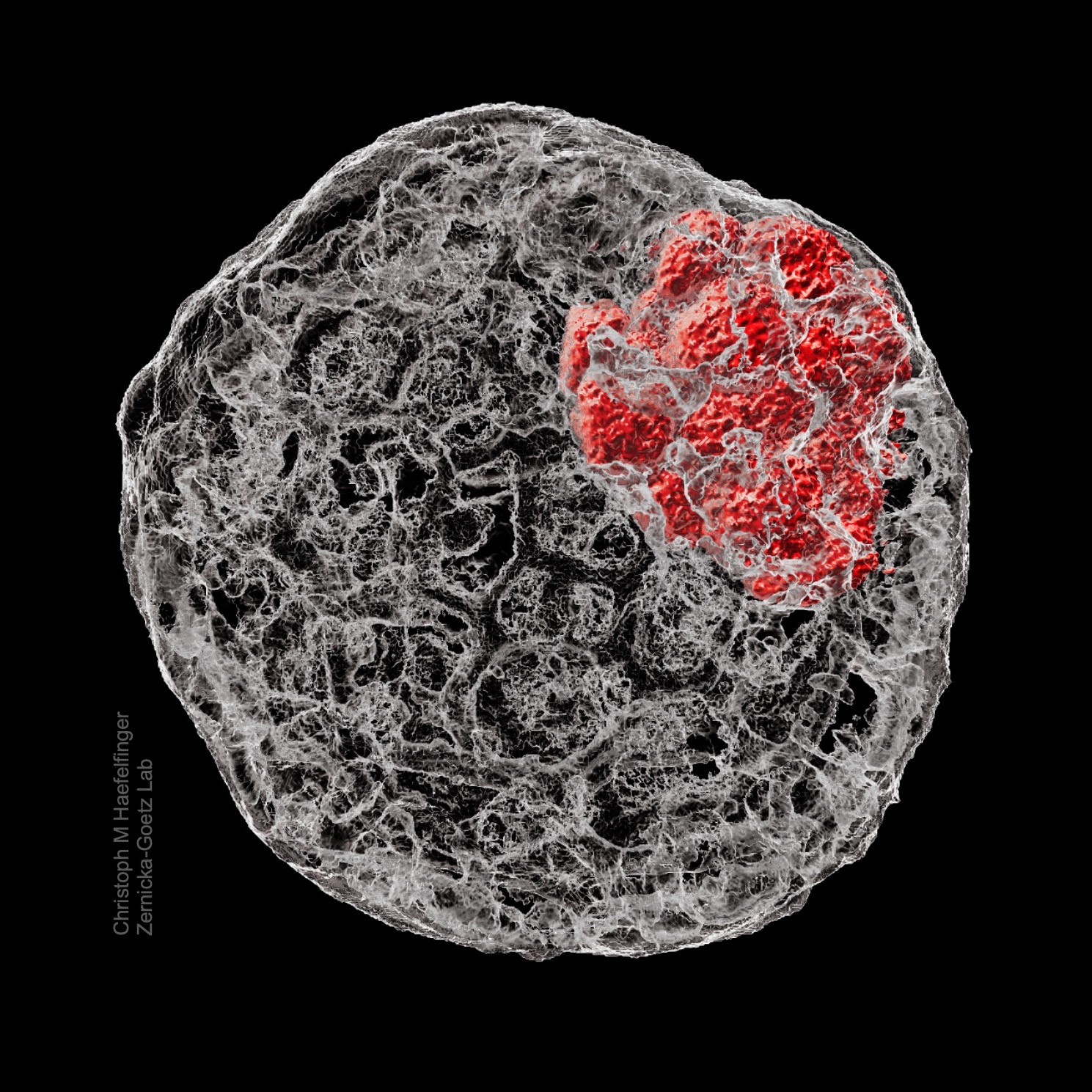
The cytoskeletal structure of preimplantation embryos demonstrated in a reconstruction of a stem cell derived mouse blastoid. After fixation, the structure was immunostained for f-actin (phalloidin, grey) and the inner cell mass (Oct4, red), then imaged.
What is your background?
I am a final year medical Student from Switzerland, currently applying for a PhD in developmental and stem cell biology in the UK. At the time I took my confocal image, I was a Summer Undergraduate Research Fellow in the laboratory of Professor Magdalena Żernicka-Goetz at the California Institute of Technology.
What are you currently researching on?
I am continuing my work at the Żernicka-Goetz lab at the University of Cambridge in parallel to clinical placements at the Universities of Cambridge and Oxford, researching preimplantation development using a mouse stem cell derived embryo model. More precisely, I aim to help solidify the scientific confidence in utilising bioengineered embryo models to study specific developmental questions.
Can you tell us more about the story behind your image ‘The backbone of stem cell derived embryos’?
To me, the image signifies the culmination of my ten week fellowship at Caltech learning the ins and outs of stem cell derived embryos, and is one of my proudest achievements. I had a truly wonderful and eye-opening experience grounded in cutting-edge research and exploring the Western USA with amazing friends and colleagues, and many of these emotions are strongly tied to my picture. I am therefore even more proud and grateful for the amazing feedback by the jury and the public – it is truly a fantastic feeling to have so many of you appreciate my image which is so close to my heart!
What is your favourite technique?
Choosing from the techniques I had the chance to learn and apply myself as an undergraduate researcher, I must go for live imaging using fluorescent reporters. I believe there are few techniques that hold the promise to not only provide quantifiable data, but to spatiotemporally visualize highly complex processes in an unparalleled beauty. Especially in the context of developmental biology, I know of no other method that could capture the dance of life in a more mesmerizing way, making it an easy choice for me.
What excites you the most in the field of developmental and stem cell biology?
I am captivated by the fact that we all originate from a single cell. To me it sometimes still is an abstract thought, and seeing an embryo develop – human, mouse, or stem cell derived – excites me every time. This enthusiasm translates into my strong curiosity for cell fate acquisition, and how it interrelates with self-organisation, spatiotemporal crosstalk, and its regulatory foundation on an omics level. I am therefore truly excited to continue researching development in my PhD.
 (1 votes)
Get involved
Create an account or log in to post your story on the Node.
Sign up for emails
Subscribe to our mailing lists.