The Maw at Etna — featured image from the Node-BSDB virtual art exhibition
Posted by the Node, on 23 November 2023
In the recent BSDB-the Node virtual art exhibition, Oliver Anderson’s ‘The Maw at Etna’ was selected as the Judges’ Choice in the ‘Scientific images’ category. We briefly caught up with Oliver to find out more about his research and the story behind the image.
Oliver Anderson (Australian Regenerative Medicine Institute)
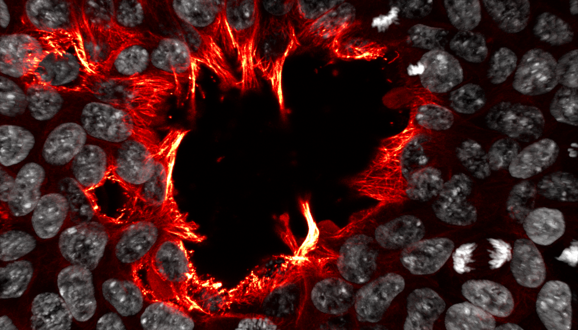
In this image, microtubules are shown in red/yellow, and nuclei in white. Cells rush to fill an opening in the colony, with their jagged flame-like microtubules roaring into the centre like the devouring forge-flames of Cyclopean Etna. (Aeneid Book VIII: Lines 416-425) Human induced pluripotent stem cells, imaged using a Zeiss LSM780 confocal microscope. Cells are labelled with DAPI (white), and immunostained for alpha-tubulin (red-yellow).
What is your background?
I did a Bachelor of Science Advanced Research (Honours) at Monash University, majoring in Genetics and Immunology. My honours project focused on modelling metabolic disease in Drosophila. I am now undertaking a PhD at the Australian Regenerative Medicine Institute in the lab of Dr Jennifer Zenker, where I am examining microtubule dynamics in human induced pluripotent stem cells (hiPSCs).
What are you currently researching on?
Currently, I am investigating and manipulating the microtubule cytoskeleton of hiPSCs in order to uncover the relationship between the structural aspects of pluripotent cells and their overall identity. Our current understanding of pluripotency is more heavily focused on genetic and metabolic aspects, and so microtubules are comparatively understudied at this stage of development.
Can you tell us more about the story behind your image ‘The Maw at Etna’?
This is one of my favourite immunostains of hiPSCs where I looked at alpha-tubulin. In this colony of hiPSCs, a hole of sorts was present in the centre of the colony, and I was struck by how the jagged intrusions looked like teeth, or even stalactites. Colouring the microtubules in red-yellow gave the appearance of fire, reminding me of Vulcan’s workshops below Etna mentioned in Aeneid Book VIII(Lines 416-425), where there’s wonderful imagery of living flames breathing through the forges, tended to by cyclops.
What is your favourite technique?
Anything that gets me on the (confocal fluorescent) microscope! Immunostaining has always given me beautiful samples that I can image slowly overnight, and techniques like transfection with fluorescent plasmids and live dyes often give fascinating live imaging data.
What excites you most in the field of developmental and stem cell biology?
There is such a huge amount we don’t understand about the beginnings of an organism’s life, and how the identities of cells transform over the course of development. Everywhere you look, there are so many questions unanswered, and to me that’s deeply exciting for the future.
 (No Ratings Yet)
Get involved
Create an account or log in to post your story on the Node.
Sign up for emails
Subscribe to our mailing lists.