Vote for a Development cover – Woods Hole – Round 3
Posted by the Node, on 19 July 2012
This week you don’t only get to decide which essay, from our competition, will appear in Development (see nominations, and the poll later today), but it’s also time to choose another cover from images from the 2011 Woods Hole embryology course. Vote in the poll below the images for the one you would like to see on the cover of Development. (Click any of the images to see a bigger version.) Poll closes on August 6, noon GMT.
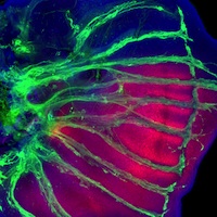
 1. 5th instar imaginal hindwing disk from the Painted Lady butterfly, Vanessa cardui. Immunostained for Engrailed in red. All nuclei are revealed by DAPI staining (blue), and trachae are shown in green. This image was taken by Alessandro Mongera, Maria Almuedo Castillo, and Jakub Sedzinski.
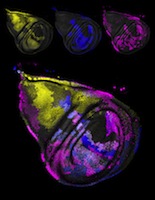
 2. 3rd instar wing disk from Drosophila melanogaster. Triple flip-out clone system (courtesy of Melanie Worley and Iswar Hariharan) was used to reveal various cell lineage clones shown in yellow, blue, and purple. All nuclei shown in gray (DAPI). This image was taken by Lynn Kee.
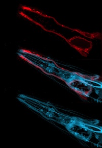
 3. Head of an adult C. elegans. DiI staining (red) reveals environmentally exposed neurons, while the JR797 GFP line allows visualization of all neurons (blue). This image was taken by Eric Brooks and John Young.
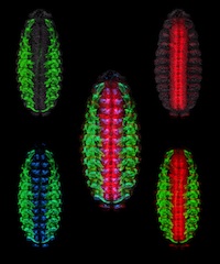
 4. Ventral view of stage 16 Drosophila melanogaster embryo immunostained for Tropomyosin (green; muscle), Pax 3/7 (blue; segmentally repeated nuclei in CNS and ectoderm), and anti-HRP (red; cell bodies and axons of the nervous system). All nuclei shown in gray (DAPI). This image was taken by Julieta María Acevedo and Lucas Leclere.
While you’re here, why not also vote for the winner of our essay competition?


 (5 votes)
I can see Kuba’s love for AB staining in that rosy wing disk
They are all very nice Confocal Z-Stack maximum intensity projections…
I chose the Drosophila embryo because that’s what my lab works on. But I must say, I really appreciated the Drosophila wing disk and C. elegans images. Due to the fact that the images use complementary colors instead of the same old primary color mashup (RGB) everyone else seems to use.
I vote for Image 4 Drosophila embryo because it is the most meaningfull and complete photo of development that I´ve ever seen.
All the photos are beautifull.
I chole drosophla embryo because is the most elegant and complete photo
Vote por la foto 4.