Vote for your favourite image in the Node–FocalPlane image competition
Posted by the Node, on 11 March 2025
To accompany the Biologists @ 100 conference, we have partnered with FocalPlane to bring to you an image competition.
We have now shortlisted 15 images, which will be presented in our gallery at Biologists @ 100 at ACC Liverpool, 24-27 March 2025, and online on the Node and FocalPlane.
Conference attendees will be able to see the images in our gallery and vote in person; for those online, you can browse through the gallery below and vote for your favourite in the poll at the bottom of this post. We’ll add up the votes from the Node, FocalPlane and our conference delegates, and the winner will be announced on Thursday 27 March.
Please vote for your favourite image at the bottom of the page. The voting will close on Wednesday 26 March 11:59pm GMT.
Thank you and good luck to the following researchers for their contributions:
Aaron Scott, Allan Carrillo-Baltodano, Andrew Octavian Sasmita, Camila Weiss, José Palma, Marina Cuenca, Çağrı Çevrim, David Grainger, Ioakeim (Makis) Ampartzidis, Julia Peloggia de Castro, Krystyna Gieniec, Lea Berg, Michael Raissig, Ludovica Altieri, Maik Bischoff, Mathieu Preußner, Min Ya and Özge Özgüç.
And a big thank you to everyone who submitted their images to the competition. There were many good quality submissions that it was very difficult to narrow down the selection!
Browse through the gallery (click to expand the images)
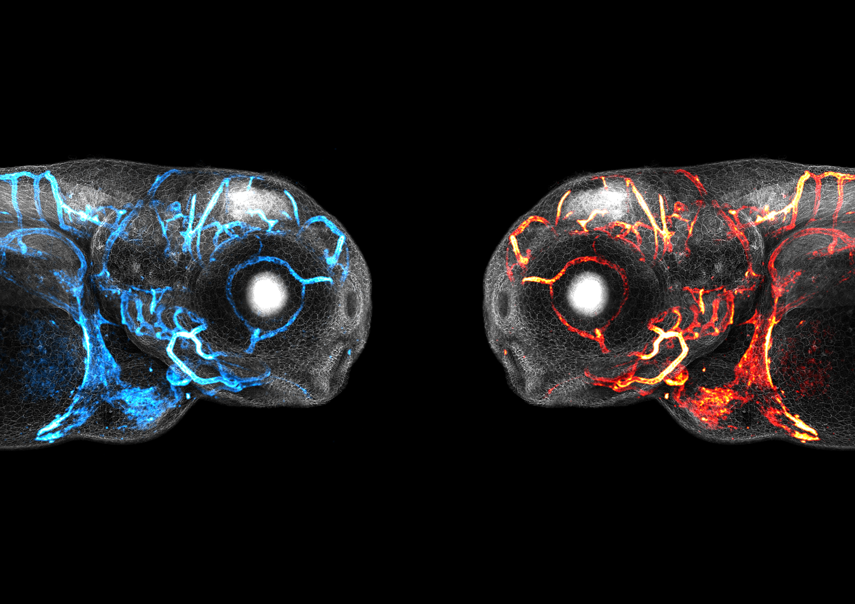
Aaron Scott
The plasma membrane of every cell in these 2-day-old larval zebrafish is fluorescently labelled and shown in grey. The endothelial cells and the blood vessels they form are shown in cyan or red. Imaged on a Leica SP8 AOBS confocal laser scanning microscope and reconstructed using ImageJ.
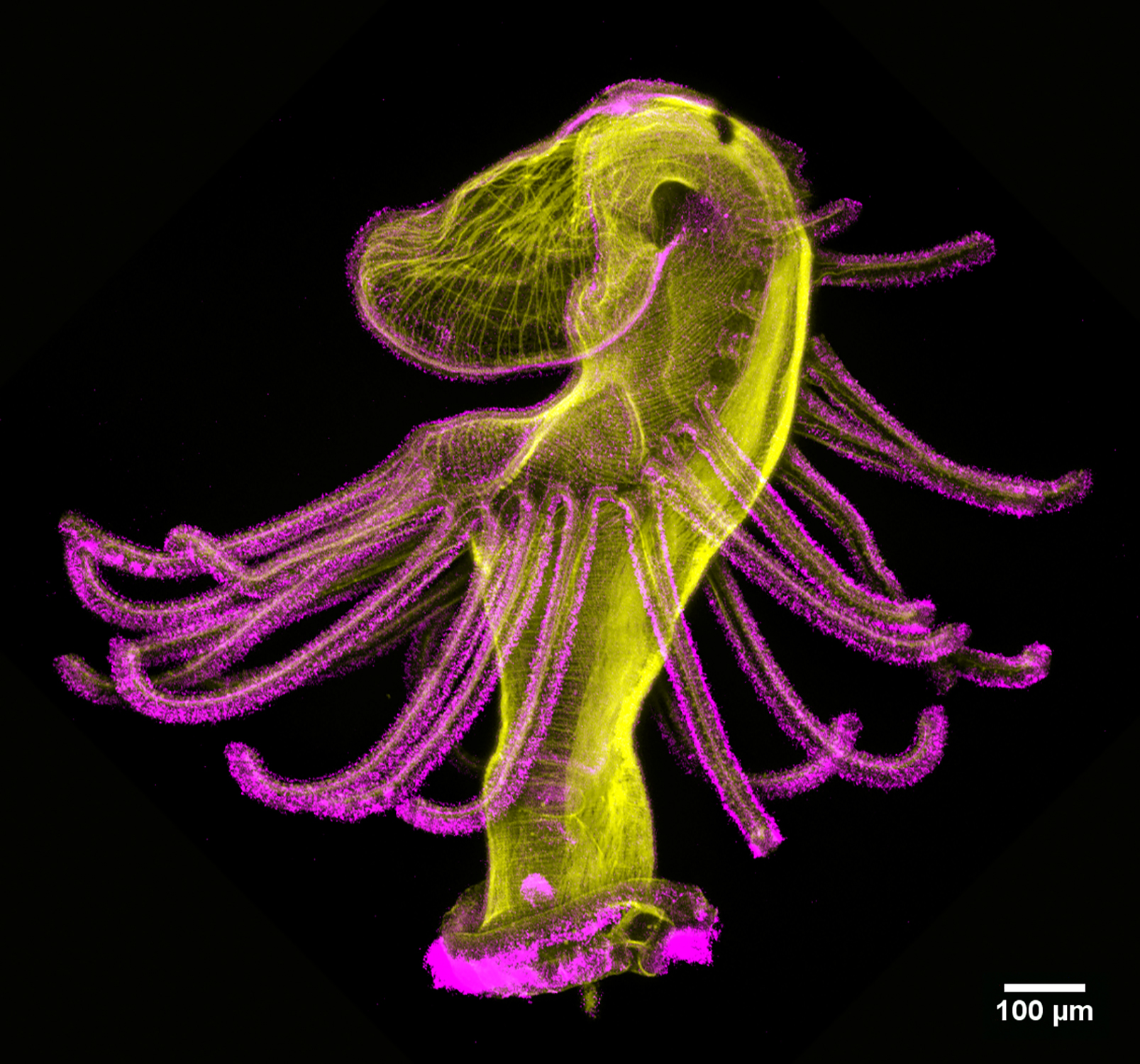
2. Dancing actinotroch
Allan Carrillo-Baltodano
Actinotroch larva of a phoronid worm with phalloidin shown in yellow and acetylated tubulin in magenta. Imaged with a Zeiss LSM 800 at 10 x magnification.
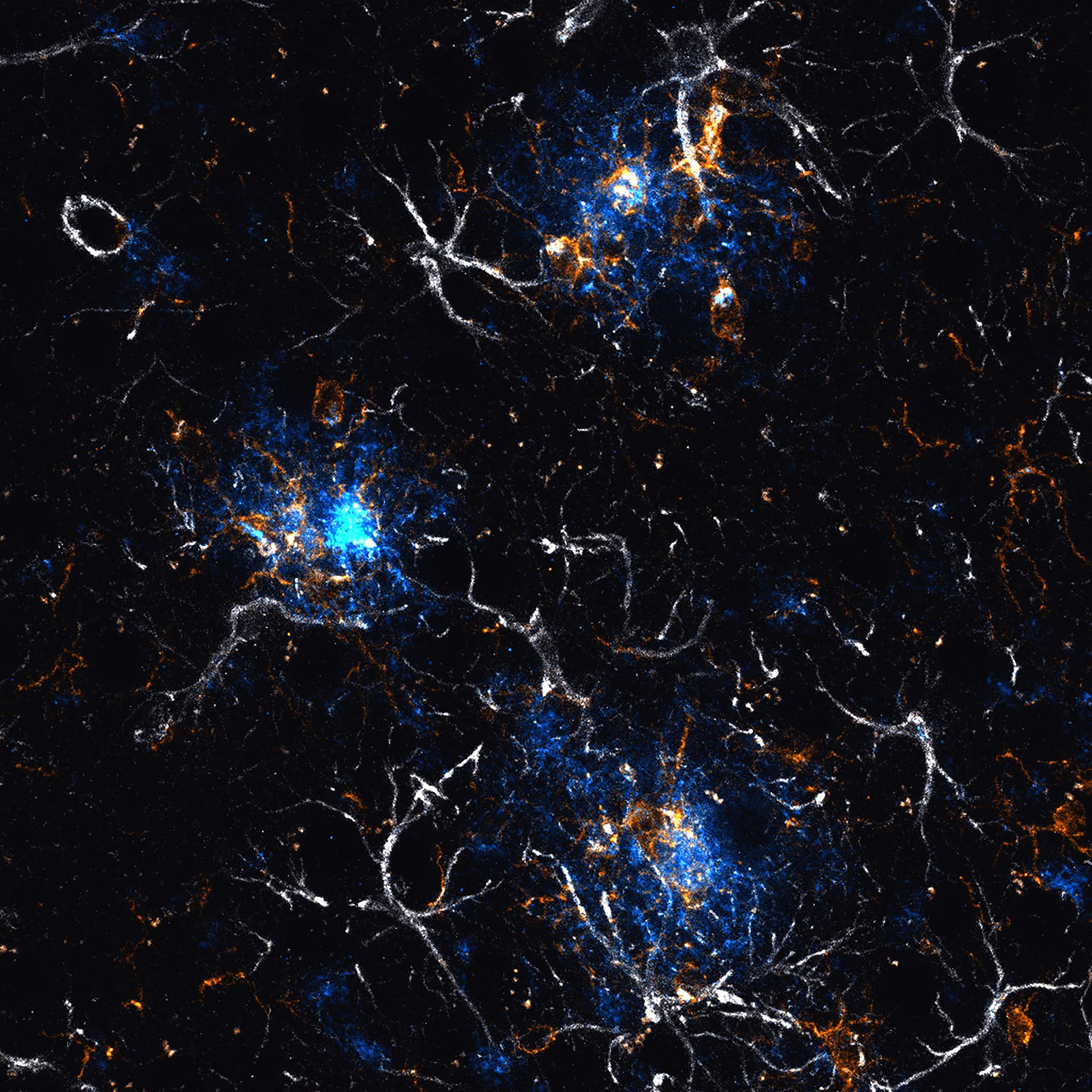
Andrew Octavian Sasmita
Confocal maximum projection image of several amyloid-β plaques (blue, 6E10) surrounded by microglia (gold, Iba1) and astrocytes (white, GFAP) in the cerebral cortex of a 6-month-old female APPNLGF mouse model of amyloidosis. Imaging was done with a Zeiss LSM 800 Airyscan confocal microscope and processed with the Zen imaging software.
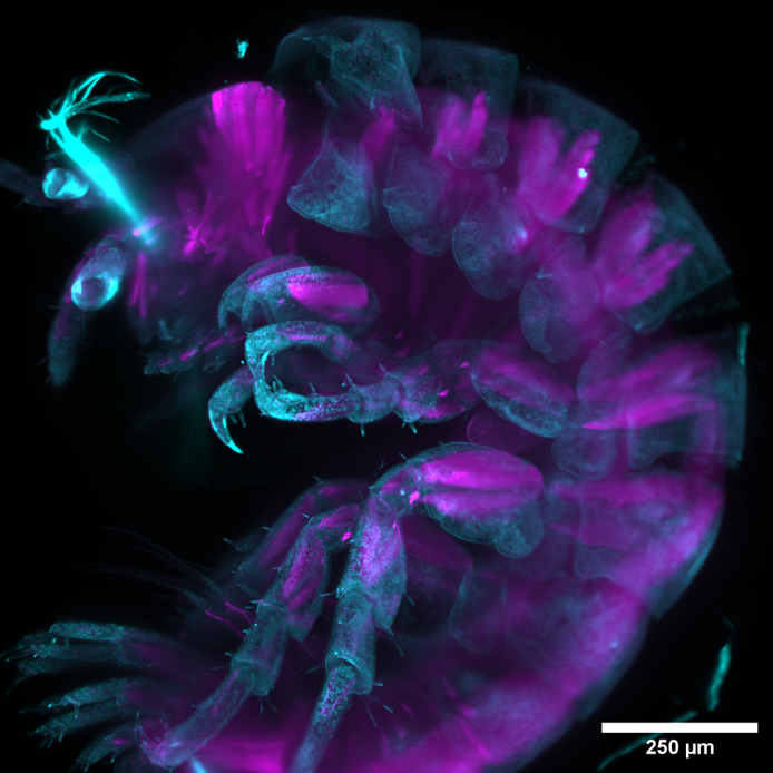
Camila Weiss, José Palma and Marina Cuenca
Lateral view of an unknown species of chilean amphipod labelled with DAPI (cyan) and phalloidin (magenta). Imaged using light-sheet imaging at the Quintay developmental biology course in 2023 and processed with Fiji.
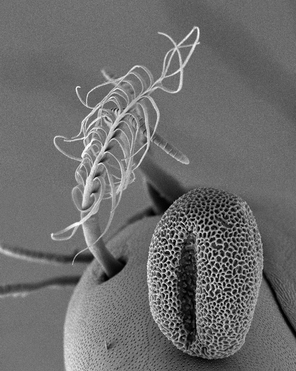
5. A beautiful contamination
Çağrı Çevrim
A scanning electron micrograph (SEM) of a Parhyale hawaiensis limb, showing an external mechanosensory organ – a plumose seta – in the background. In the foreground, a pollen grain, possibly from a Platanus tree, has contaminated the sample.
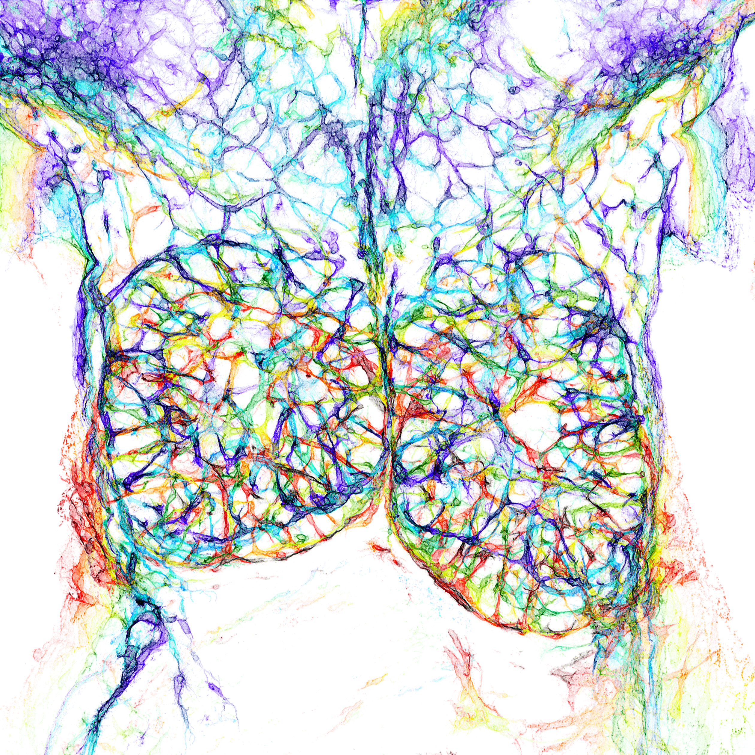
6. Thymus in the spotlight
David Grainger
A colour-coded depth projection of the blood endothelial cells of the E14.5 mouse embryonic thymus and surrounding structures. A 200 μm thick vibratome section was immunostained for endomucin and imaged on a Zeiss LSM980 confocal microscope and depth-encoded using a rainbow LUT before performing a maximum intensity projection.
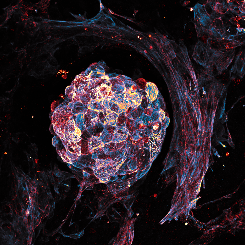
7. Beat
Ioakeim (Makis) Ampartzidis
Mature beating cardiomyocyte cell cluster from human induced pluripotent stem cells. Cells were grown for a total of 20 days and stained positive for cardiac troponin (Hot Blue) and actinin (Hot Red) markers. The image was acquired at the Veneto Institute of Molecular Medicine (VIMM), in Nicola Elvassore’s lab, using an upright LSM900 ZEISS microscope and LUTs adjusted using Fiji software.
8. Who’s active?
Julia Peloggia de Castro
The image depicts a zebrafish embryo at 9 hours post-fertilisation on a lateral view. Cells are stained with MitoTracker, which labels active mitochondria, and cell membranes are labelled in cyan with a EGFP transgenic membrane tag. Image was taken using a 20x objective on a spinning disk confocal microscope.

Krystyna Gieniec
2D culture of mouse mammary fibroblasts stained for Acta2 (magenta) and Vimentin (gold), with some contaminating epithelial cells stained for pan-Cytokeratin (cyan). Image acquired using a Leica Stellaris 8 confocal microscope.
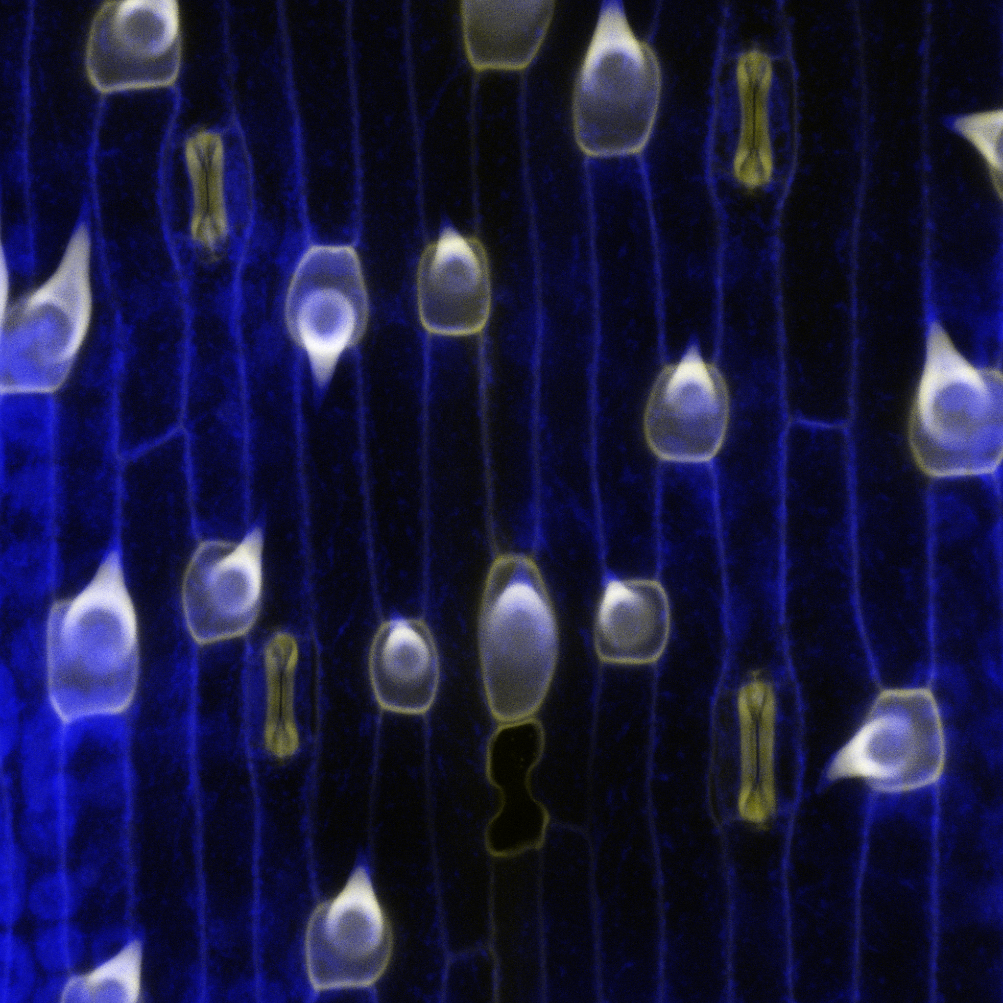
10. The plant-atmosphere interface that feeds the world
Lea Berg and Michael Raissig
Mature epidermal cell types in a grass leaf of the emerging developmental model system Brachypodium distachyon. Cell outlines are blue, which is plant cell wall UV-autofluorescence. In yellow is stained lignin, a secondary cell wall modification that can be found in the hair cells (‘shark-tooth’-shaped) and the stomatal guard cells (‘dumbbell’-shaped). Imaged by confocal microscopy and processed in Fiji.
11. Plenty of fish in the sea
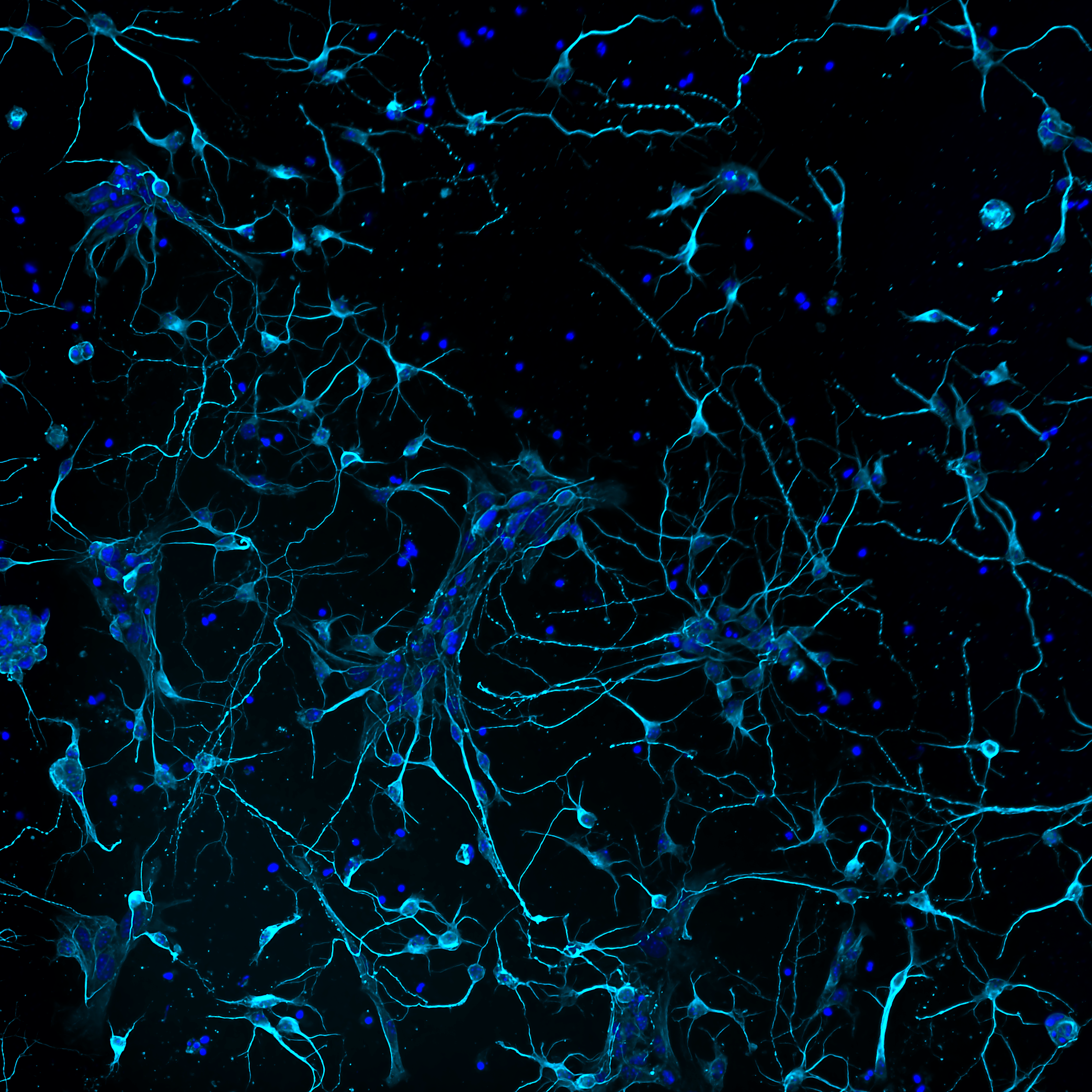
Ludovica Altieri
Murine primary cortical neurons developing interconnections, stained with neuronal tubulin (cyan) and DAPI (blue). Imaged on a Nikon microscope implemented with a CrestOptics confocal spinning disk module with post-processing using NIS Elements AR by Nikon. Acquired at the IBPM Institute of Molecular Biology and Pathology – CNR National Research Council of Italy, c/o Department of Biology and Biotechnology “Charles Darwin” – Sapienza University of Rome.

12. Invisible architects
Maik Bischoff
Drosophila hydei testis musculature stained with phalloidin to label F-actin (blue), anti-N-Cadherin (orange/gold) to mark cell-cell junctions between muscle cells and DAPI to stain nuclei (purple). Autofluorescence (orange/gold) makes the trachea visible. Imaged with a Zeiss 980 confocal microscope with Airyscan 2. The image was processed in Zen Blue, and LUTs (by KTZ) applied in Fiji with further modifications in Photoshop.
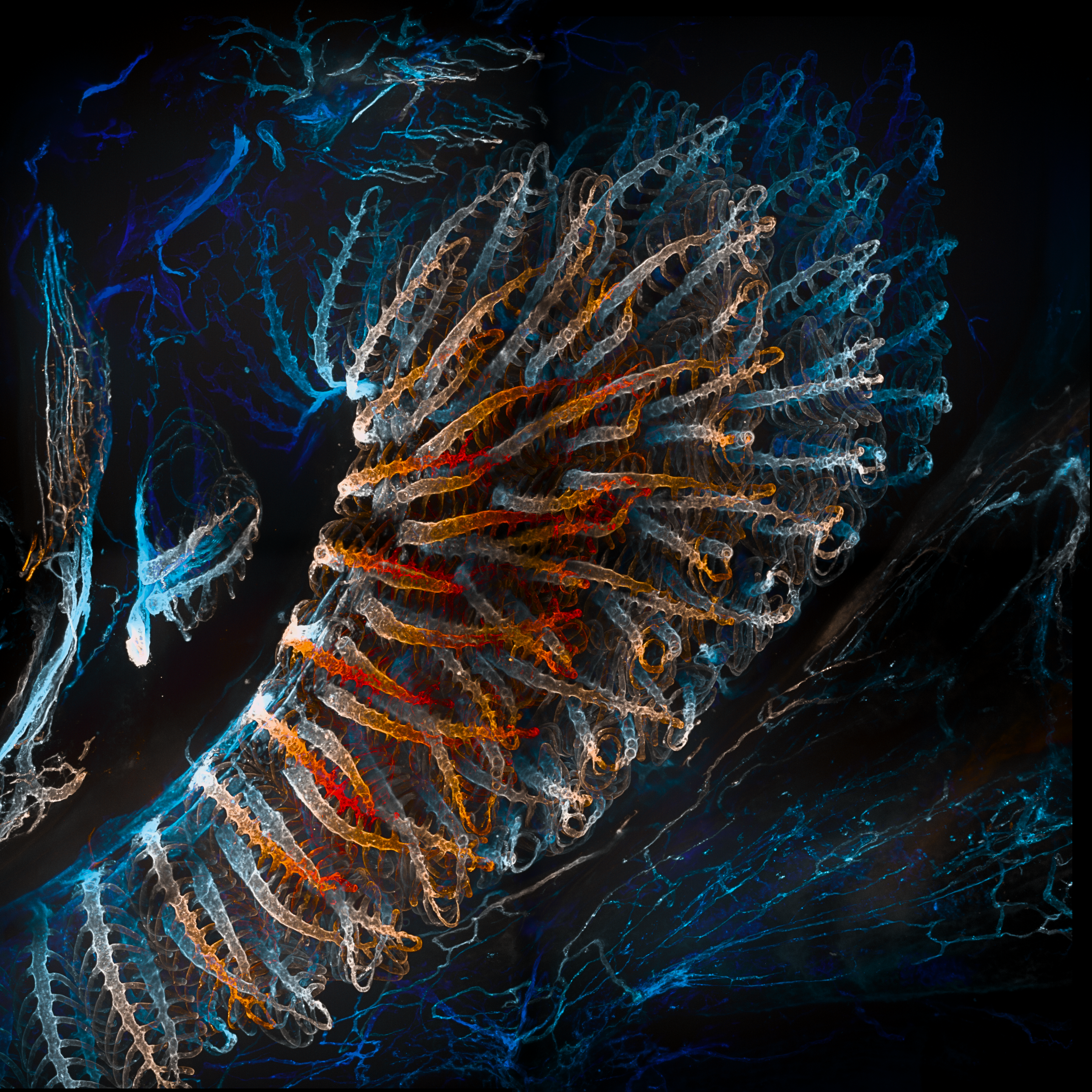
Mathieu Preußner
Lateral view of the overlying gill arches in 1-month-old Danio rerio expressing endothelial kdrl:mCherry. Clarity-based tissue clearing of the sample enabled comprehensive image acquisition using a Nikon Ti spinning disk system. In ImageJ, the hyperstack was modified using a temporally colour-coded lookup table.
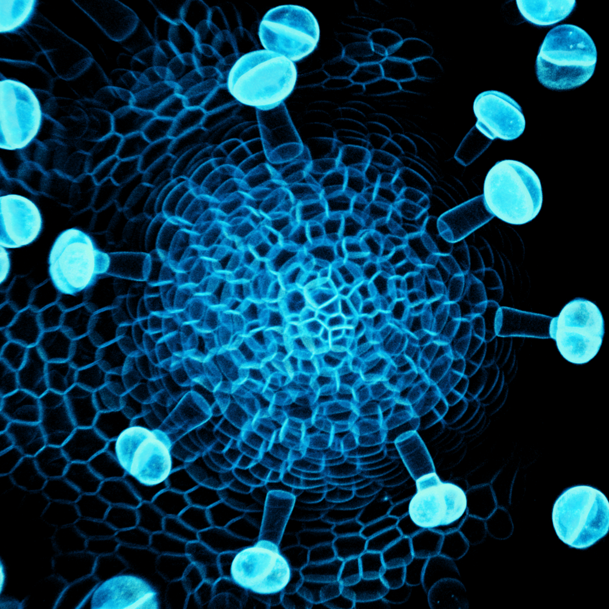
Min Ya
Maximum projection of confocal stacks of a mutant Mimulus parishii shoot apex with cells labelled with a plasma membrane marker. The shoot apices of this plant can grow but are unable to produce any organs, resulting in a phenotype that resembles a pin.
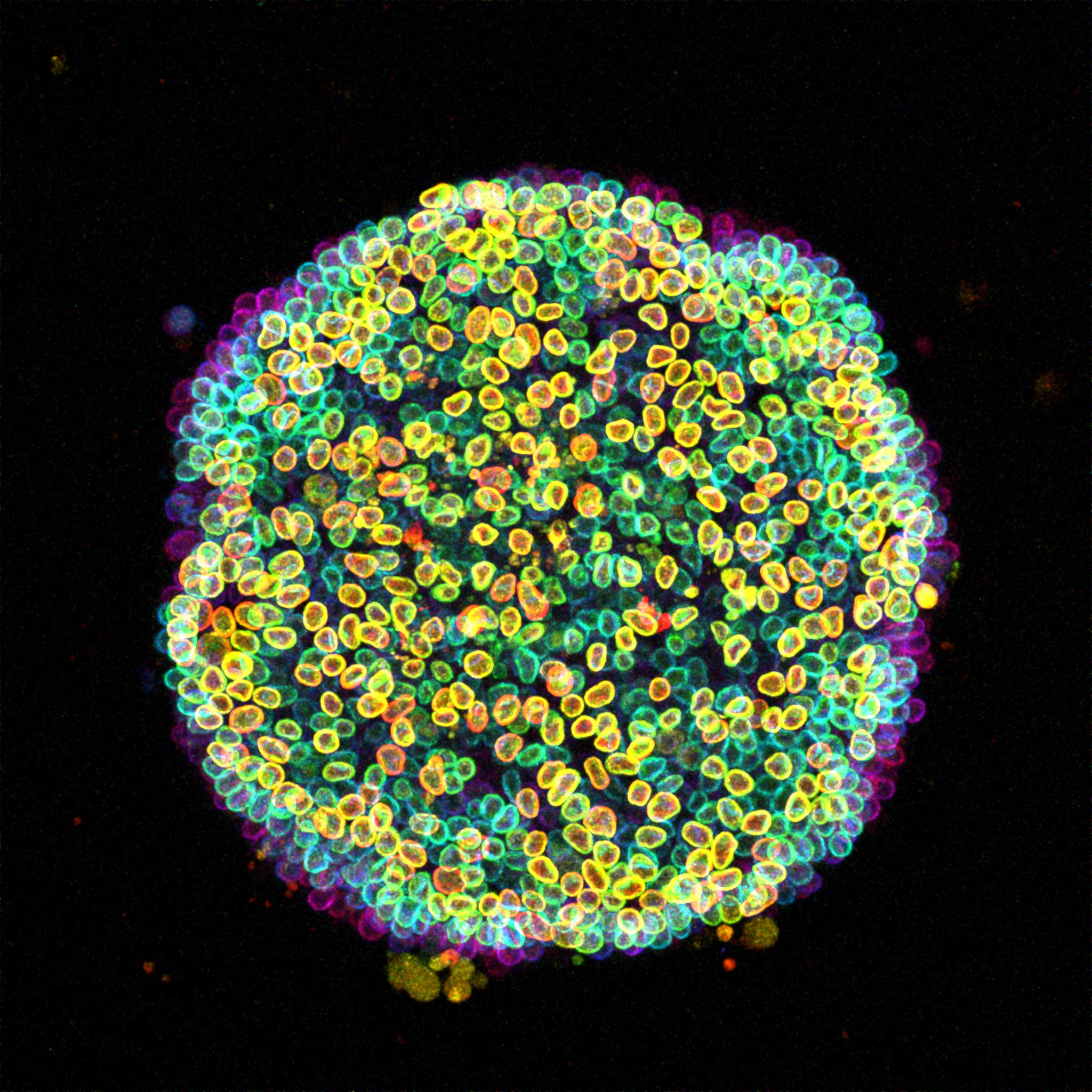
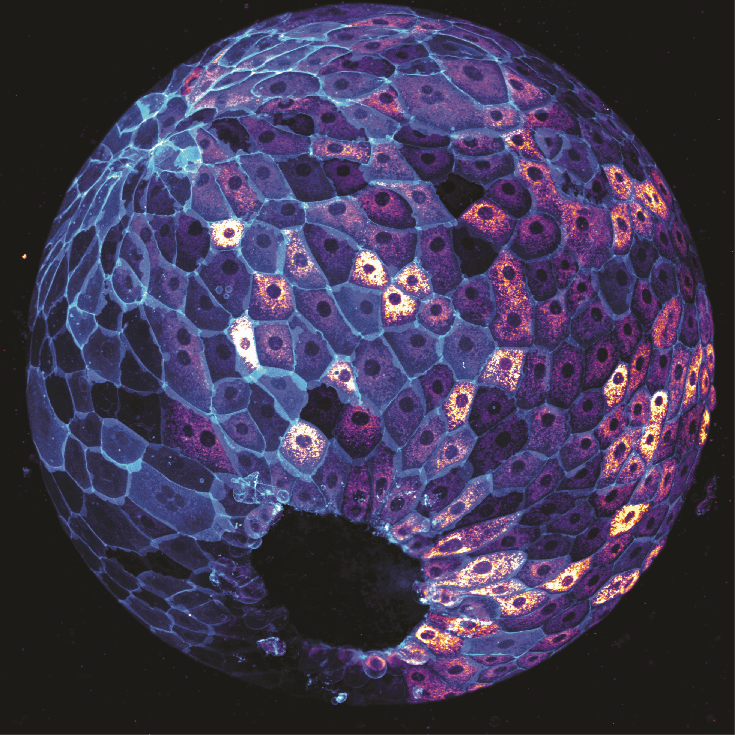
Özge Özgüç
A ‘Cell-estial bloom’ of human induced pluripotent stem cells (hiPSCs) flourishes on a micropatterned island. This image presents a colony of live hiPSCs, with fluorescently labelled Lamin B delineating the nuclear lamina within each cell. Acquired with a Zeiss LSM 880 Airyscan microscope, this maximum intensity projection is enhanced with depth-coded coloring to reveal the captivating three-dimensional landscape.
Voting is now closed. Thank you to everyone who voted!
 (172 votes)
35 thoughts on “Vote for your favourite image in the Node–FocalPlane image competition”
Leave a Reply
Get involved
Create an account or log in to post your story on the Node.
Sign up for emails
Subscribe to our mailing lists.
Very nice
Amazing images!
Nr. 13
they were all nice images! Congrats to all!
👏
Çok güzel 👍🏼
Beautiful 😍
Nr: 15
Bright colors
Fascinating 💫
❤️
Marvelous 💜
Çok güzel 💕
What an awe-inducing shot!!
Pure beauty
Speechles!
belle photo; félicitations
Wonderfull
Very good🙏🏻🙏🏻🙏🏻👏👏👏
Artistic science. ♥️
👍
🩷
👍
Marvelous👏👏
👏👌🏻
İ love that
Harika, çok güzel görüntüler. Görüntüler için çaba harcayan, emek koyan herkese teşekkür ederim.
👏👏
All images are beautiful!
All imagens are beautiful!!!
Todas lindas e sutis!!
Muitas imagens me tocaram pela beleza!
Parabéns aos participantes!
Sei do grande trabalho de todos.
Beautiful images, compliments to everybody!!!
Congratulations
Dear Övgü,
Congratulations on your achievement! Your hard work and dedication have truly paid off. Wishing you continued success in all your future endeavors. May this be just the beginning of many more accomplishments to come!
Özgü I really liked your work. Congratulations on your achievement and wishing you both continued success in the future.
We support you, Özge!