Building strength and stability: assembly of tendon-bone attachment
Posted by Erin M Campbell, on 12 July 2013
 I appreciate my health and body, but I admit that there are times when I take all of the amazing biology inside my body for granted. My bones and muscles help me easily type this sentence, but the coordination that takes place to form the musculoskeletal system is far from simple. Today’s image is from a recent Development paper that finds the source of cells that form the tendon-bone attachment unit.
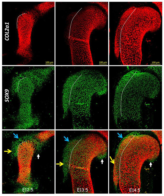
Tendons, muscles, and bones all work together to provide us with strength, stability, and mobility. Tendons connect muscles to the bone, and can withstand tension to provide additional stability. The tendon-bone attachment unit is a complex structure where the tip of the tendon is inserted into specific sites on the bone called eminences, and its formation during development is not completely understood. A recent paper identifies the source of progenitor cells that make up the bone eminence. Blitz and colleagues found that bone eminence progenitor cells are not descendants of cartilage-forming chondrocytes, which originate from mesenchymal stem cells in the lateral plate mesoderm. Instead, the tendon-bone attachment unit cells come from an external pool of progenitor cells that express both Sox9 and Scx. The eminences develop after early cartilage tissue has been formed, and are modularly added onto the bone. The specification of these progenitors depends on TGFβ signaling, and the differentiation of these progenitors into eminence cells depends on BMP4 signaling. In the images above, sections of the humerus bone (red shows cartilage, collagen II) in mice at different developmental stages show an external pool of Sox9-positive, Col2A1-negative cells (green, arrows) that differentiates into the tendon-bone attachment unit. These domains later differentiated and contained collagen II (right column).
For a more general description of this image, see my imaging blog within EuroStemCell, the European stem cell portal.
Personal Note: This will be my last post on The Node and EuroStemCell for a few months, as I await the August arrival of my second daughter. I’ll be keeping up with exciting stem cell discoveries when not totally sleep-deprived, and I look forward to posting more images soon! Thanks for reading!
![]() Einat Blitz, Amnon Sharir, Haruhiko Akiyama, & Elazar Zelzer (2013). Tendon-bone attachment unit is formed modularly by a distinct pool of Scx- and Sox9-positive progenitors Development, 140 (14), 2680-2690 DOI: 10.1242/dev.093906
Einat Blitz, Amnon Sharir, Haruhiko Akiyama, & Elazar Zelzer (2013). Tendon-bone attachment unit is formed modularly by a distinct pool of Scx- and Sox9-positive progenitors Development, 140 (14), 2680-2690 DOI: 10.1242/dev.093906
 (7 votes)
Get involved
Create an account or log in to post your story on the Node.
Sign up for emails
Subscribe to our mailing lists.