2nd European Developmental Biology Congress (EDBC 2023) – The Oxford Panorama
Posted by Nawseen Tarannum, on 9 January 2024
Nawseen Tarannum, postdoc, University of Manchester
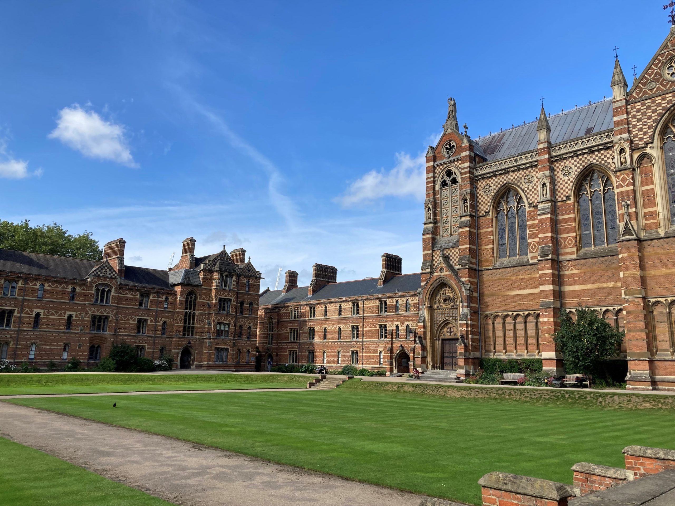
While conference hunting in early 2023, I saw an advertisement for the 2nd EDBC on the BSDB website. Given my love for cell and developmental biology, wanting to attend it was a no-brainer decision. Held from 25th-28th September 2023 in Oxford, the conference attracted developmental biologists from across Europe. Packed with a collection of great talks, posters and social events, this conference was unique for the science and how it was organised. In addition to Oxford, there were two hubs – Paris and Barcelona – from where talks were live-streamed and intertwined with the Oxford schedule. I’m not sure what technology gods the organisers and the AV team prayed to, but they managed to pull off the least technologically disruptive hybrid conference I’ve been to. Truly impressive. Now let’s turn to the real showstopper, the science.
Pilot
Season two of the conference started in the afternoon with an opening address from Paul Martin (BSDB chair). He promised fantastic talks prioritising early-career group leaders and researchers and the conference programme indeed delivered on this promise. He also introduced the winner of the Cheryll Tickle medal, a mid-career female scientist who has made outstanding contributions to the field. This year’s medal was awarded to Madeline Lancaster. She took us on a trip through her PhD and postdoc days before focusing on her lab’s interest in understanding how the human brain develops compared to other primates. Her work has identified that the tissue architecture of the developing brain impacts cell fate, with temporal progression from progenitors to neurons depending on the correct spatial positioning of cells (1).
Madeline’s presentation paved the way for other talks that afternoon that were based on “patterning”. During this session, I listened to Teresa Rayon talk about discrepancies in the developmental timescale of neural tube ventral patterning in mice and humans. She showed that although the process is conserved between both species, increased protein stability in human cells may explain our slower development (2). Jacqueline Tabler discussed skull morphogenesis in the context of a noncanonical form of cell motility. She showed that a collagen gradient drives osteoblast movement, divisions and differentiation towards a softer matrix with feedback between the stiffness gradient and cell fate controlling bone size (3). Markéta Kaucká Petersen elaborated on how her lab outlines the blueprint of cellular heterogeneity underlying craniofacial morphogenesis using single-cell genomics and transcriptomics.
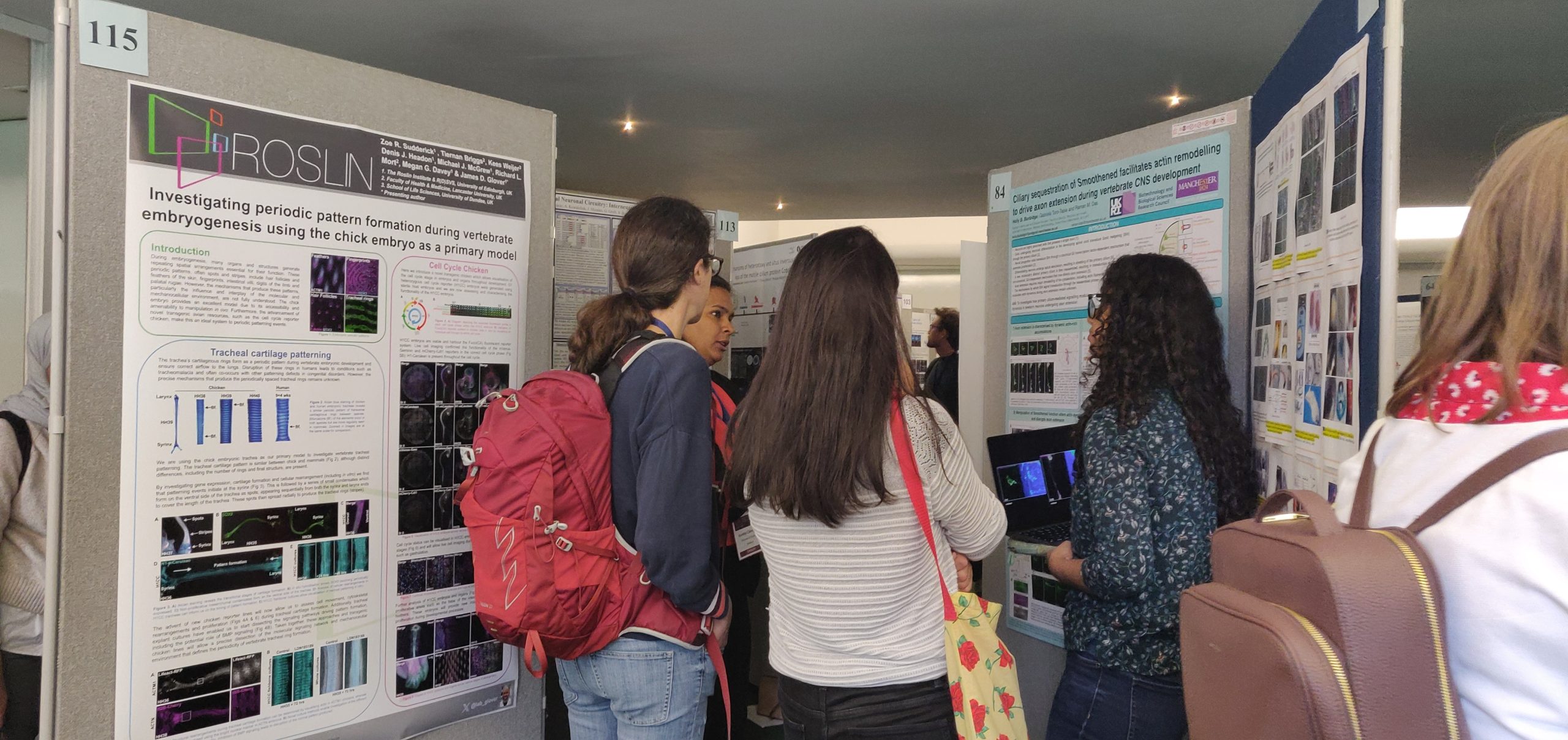
The relatively short first day was brought to a close with a poster session featuring a virtual reality experience, designed by Shaping Destiny, involving dance forms that depict embryonic growth. Unfortunately, I missed this as I got too immersed in the poster session (recruiting people to my poster), but those who experienced it said it was good fun. Hopefully, I’ll be able to try it at the next EDBC (or another BSDB conference)!
Episode 2
After a well-needed sleep, day two began with some morning Tai chi led by in-house Shifu Paul Martin. Those of us who braved the drizzly outdoors witnessed the Shifu very meticulously yet hilariously demonstrate how we could potentially defend ourselves. I, however, was embarrassingly hopeless, and then it was time to join the first session of the day to remind myself of biology which I feel I am marginally better at compared to Tai chi.
Day two was all about the hybrid format with interdigitating talks between Oxford and Paris. We started with the “morphogenesis” session, where Magali Suzanne described how apoptotic cells exert apicobasal forces on the neighbouring tissue and actively contribute to neural tube bending (4). Similar forces are also generated during epithelial-to-mesenchymal transition (EMT) in Drosophila (5). I also listened to Diana Pinheiro explain how a gradient of Nodal signalling fractionates the zebrafish mesendoderm into highly protrusive leader cells and less protrusive followers. The leader cells readily undergo internalisation while they pull the followers inwards in a mechanism that preserves mesoderm patterning (6). An interesting talk by Thibaut Brunet introduced the multicellular choanoflagellate, Choanoeca flexa. These remarkable organisms form multicellular sheets of polarised cells reminiscent of an epithelial monolayer (7) and reversibly transition between the multicellular colony state and unicellular dormant cysts depending on environmental conditions. The session ended with a visually stunning talk from Kate McDole who has developed an advanced light sheet microscope to investigate mouse organ development. By applying machine learning to the images, her lab follows the journey of individual cells up to organogenesis (8). Interestingly, this microscope can track the embryo’s position at complex developmental stages and adapt for optimal imaging without requiring manual input. How I wish I could use this fancy smart scope for my experiments!
After a morning of fascinating talks, the afternoon provided the much-awaited opportunity to explore beautiful Oxford. Some of us visited the botanical garden while others went punting. I decided to see some pretty flowers and when I returned in two hours, my phone was brimming with photos of colourful petals while my airways were revved up with pollen. Thankfully, I did not let out one of my signature deafening sneezes to disrupt the upcoming talks.
The afternoon was exclusively dedicated to talks from Paris with the theme “Gene regulation”. The opening talk by Claire Rougeulle showed that X chromosome inactivation and dampening are regulated by the same molecular players during early development in females (9). Nicola Festuccia then highlighted the transcription factor Nr5a2 as a master regulator of gene expression coordinating proliferation and genome stability during preimplantation in mice (10). At the end of the talks, we were treated to a grand dinner at the Hogwarts-esque dining hall of Keble College. With our stomach content, we celebrated the success of some ace researchers. Jonathan Slack was awarded the Wolpert medal recognising his impact through public outreach and teaching especially as he has written multiple books for scientific and non-scientific audiences. The Waddington medal recipient, whose identity was kept top secret until the award, was Marysia Placzek. The medal symbolised her major contributions to developmental biology, which became apparent as she took us through her research journey. The thousands of embryos she has dissected throughout her career were enough grounds to earn her the medal although her research on understanding patterning of the vertebrate nervous system was probably what did the trick. Alongside her career trajectory, it was amazing to see how she balanced her family life, a quality I find inspiring being a woman in science. To top it all off, the evening ended with a Hollywood-style trailer of the upcoming BSDB film that collated the diverse scenes of developmental biology in the UK.
Episode 3
The morning of day three was designed around “Concepts and theories in developmental biology”. Inaugurating the session, Jeremy Green delved into the theories of pattern formation based on the “reaction-diffusion model” and “positional information” theory (11). He also proposed two new ideas – mechanics and an omics approach to morphogenesis – in symmetry breaking. In a related aspect, Berta Verd showed that mathematical models of pattern formation should implement cell movements that are ignored in such models. Her research has established, for the first time, a framework to reverse-engineer gene regulatory networks (GRNs) of pattern formation in tissues under robust morphogenetic cell rearrangements (12). I also listened to James DiFrisco discuss the conundrum of how generalised principles can be extracted from diverse biological systems and applied across phylogenies. He suggested that combining mathematical modelling and our knowledge of evolutionarily conserved mechanisms may help address such complexities. Finally, Ruth Baker demonstrated that calibrating a mathematical model to high throughput experimental data from scratch assays can be used to infer mechanistic details underlying wound closure (13). Fitting right in with the session’s theme, the Beddington medal was awarded to Rasa Elmentaite for her fantastic PhD thesis investigating human intestinal development using single-cell RNA sequencing and spatial transcriptomics (14, 15).
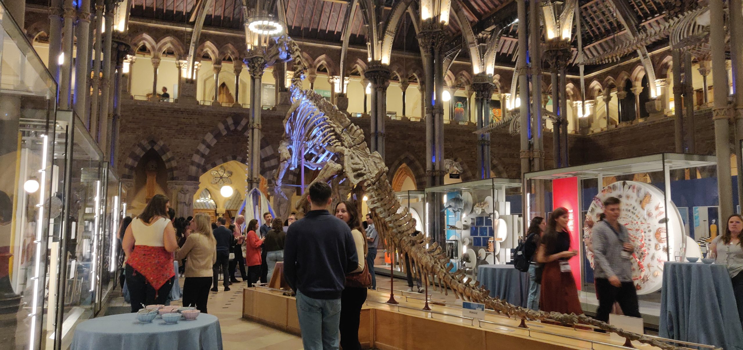
The afternoon was Barcelona’s moment in the sun (quite literally), similar to the format of day two. With quite a vast genre encompassing “Dynamics, mechanics and evolution”, this session was packed with interesting talks. Alejo Rodriguez-Fraticelli outlined how combining single-cell genomics and lineage tracing can be used to understand cellular heterogeneity and its consequences during development, ageing and cancer (16, 17). Yanlan Mao spoke about how tissues maintain their shape during repair and homeostasis. Focusing on wound closure in the Drosophila wing disc, she showed that tissue fluidity at the wound edge helps repair (18) with mechanical force-mediated cell shape changes contributing to the process. Next, Elvan Böke probed the question, “How do oocytes remain healthy for decades?” to which a part of the answer could be due to the ability of dormant oocytes to keep two detrimental factors in check – the production of reactive oxygen species (19) and the accumulation of protein aggregates in the cytosol. In his talk, Cristian Cañestro revealed how the tunicate, Oikopleura dioica, can be used to study gene loss and the deconstruction of GRNs to understand evolutionary diversity (20). Lastly, Antonio Scialdone showed that loser cells possessing mitochondrial defects tend to be eliminated in the mouse embryonic epiblast with cell competition selecting for optimal mitochondrial function before gastrulation (21). After a day of back-to-back talks, the evening was party time with the dinosaurs. Drinks in hand, we explored the quirky Pitts River Museum before ending up at the Museum of Natural History, our final stop for the night. With great food, drinks, music and dancing I unwinded in the revelry. If only the dinosaurs had come alive, it would have been a perfect re-enactment of Night at the Museum…
Season finale
After partying (and one too many drinks), it was nothing short of a miracle that I made it to the first talk the following morning. It was presented by Julien Leclercq who was also awarded the Thesis Prize from the French Society for Developmental Biology for his outstanding PhD research identifying the genetic mechanisms that regulate eye formation in different morphotypes of the fish Astyanax mexicanus (22). Julien’s presentation was followed by the final instalment of talks revolving around “Regeneration, Disease and Ageing”, where Mathilda Mommersteeg challenged the current notion that oxidative phosphorylation inhibits regeneration. She highlighted that oxidative phosphorylation is required for the re-differentiation of cardiomyocytes and the long-term regeneration of the zebrafish heart. Stephanie Ellis then focused on how cell competition maintains tissue structure during mouse skin development. She outlined two sequential mechanisms – (a) the elimination and engulfment of loser cells by winners and (b) the expulsion of losers from the basal layer by differentiation (23). In the penultimate talk of the conference, Daria Siekhaus demonstrated that BMP signalling specifies the fate of leader macrophages that then infiltrate the Drosophila embryo to regulate homeostasis. Lastly, the closing keynote was delivered by Angela Nieto who talked about different programmes of EMT concerning three scenarios – development, fibrosis, and cancer. She discussed the commonalities and differences between these EMT forms and how they can maintain or breach epithelial homeostasis.
After four days of excellent talks, fruitful discussions and making new science buddies, it was time to return to life as usual. While on my train to Manchester, I felt grateful to have attended the EDBC. It not only provided important feedback for my work but also instilled in me new ideas that I could explore. I learned about aspects of developmental biology that I wasn’t familiar with before, helping broaden my outlook on the field and making me realise the scale of the versatile things that are indeed possible. While writing this report, I eagerly look forward to the next one. Here’s to EDBC 2027!
Head over to read Tanya Foley’s meeting report from the Paris hub’s perspective!
References
1. Chiaradia I, Imaz-Rosshandler I, Nilges BS, Boulanger J, Pellegrini L, Das R, et al. Tissue morphology influences the temporal program of human brain organoid development. Cell Stem Cell. 2023;30(10):1351-67.e10.
2. Rayon T, Stamataki D, Perez-Carrasco R, Garcia-Perez L, Barrington C, Melchionda M, et al. Species-specific pace of development is associated with differences in protein stability. Science. 2020;369(6510).
3. Dang Y, Lattner J, Lahola-Chomiak AA, Afonso DA, Taubenberger A, Ulbricht E, et al. Self-propagating wave drives noncanonical antidurotaxis of skull bones in vivo. bioRxiv. 2023:2023.07.10.547677.
4. Roellig D, Theis S, Proag A, Allio G, Bénazéraf B, Gros J, Suzanne M. Force-generating apoptotic cells orchestrate avian neural tube bending. Dev Cell. 2022;57(6):707-18.e6.
5. Gracia M, Theis S, Proag A, Gay G, Benassayag C, Suzanne M. Mechanical impact of epithelial-mesenchymal transition on epithelial morphogenesis in Drosophila. Nat Commun. 2019;10(1):2951.
6. Pinheiro D, Kardos R, Hannezo É, Heisenberg C-P. Morphogen gradient orchestrates pattern-preserving tissue morphogenesis via motility-driven unjamming. Nature Physics. 2022;18(12):1482-93.
7. Brunet T, Larson BT, Linden TA, Vermeij MJA, McDonald K, King N. Light-regulated collective contractility in a multicellular choanoflagellate. Science. 2019;366(6463):326-34.
8. McDole K, Guignard L, Amat F, Berger A, Malandain G, Royer LA, et al. In Toto Imaging and Reconstruction of Post-Implantation Mouse Development at the Single-Cell Level. Cell. 2018;175(3):859-76.e33.
9. Alfeghaly C, Castel G, Cazottes E, Moscatelli M, Moinard E, Casanova M, et al. XIST dampens X chromosome activity in a SPEN-dependent manner during early human development. bioRxiv. 2023:2023.10.19.563078.
10. Festuccia N, Vandormael-Pournin S, Chervova A, Geiselman A, Langa-Vives F, Coux R-X, et al. Nr5a2 is essential for morula development. bioRxiv. 2023:2023.01.16.524255.
11. Green JB, Sharpe J. Positional information and reaction-diffusion: two big ideas in developmental biology combine. Development. 2015;142(7):1203-11.
12. Spiess K, Fulton T, Hwang S, Toh K, Saunders D, Paige B, et al. Approximated Gene Expression Trajectories (AGETs) for Gene Regulatory Network Inference on Cell Tracks. bioRxiv. 2022:2022.01.12.476060.
13. Martina Perez S, Sailem H, Baker RE. Efficient Bayesian inference for mechanistic modelling with high-throughput data. PLoS Comput Biol. 2022;18(6):e1010191.
14. Elmentaite R, Kumasaka N, Roberts K, Fleming A, Dann E, King HW, et al. Cells of the human intestinal tract mapped across space and time. Nature. 2021;597(7875):250-5.
15. Elmentaite R, Ross ADB, Roberts K, James KR, Ortmann D, Gomes T, et al. Single-Cell Sequencing of Developing Human Gut Reveals Transcriptional Links to Childhood Crohn’s Disease. Dev Cell. 2020;55(6):771-83.e5.
16. Rodriguez-Fraticelli AE, Wolock SL, Weinreb CS, Panero R, Patel SH, Jankovic M, et al. Clonal analysis of lineage fate in native haematopoiesis. Nature. 2018;553(7687):212-6.
17. Weinreb C, Rodriguez-Fraticelli A, Camargo FD, Klein AM. Lineage tracing on transcriptional landscapes links state to fate during differentiation. Science. 2020;367(6479).
18. Tetley RJ, Staddon MF, Heller D, Hoppe A, Banerjee S, Mao Y. Tissue Fluidity Promotes Epithelial Wound Healing. Nat Phys. 2019;15(11):1195-203.
19. Rodríguez-Nuevo A, Torres-Sanchez A, Duran JM, De Guirior C, Martínez-Zamora MA, Böke E. Oocytes maintain ROS-free mitochondrial metabolism by suppressing complex I. Nature. 2022;607(7920):756-61.
20. Ferrández-Roldán A, Fabregà-Torrus M, Sánchez-Serna G, Duran-Bello E, Joaquín-Lluís M, Bujosa P, et al. Cardiopharyngeal deconstruction and ancestral tunicate sessility. Nature. 2021;599(7885):431-5.
21. Lima A, Lubatti G, Burgstaller J, Hu D, Green AP, Di Gregorio A, et al. Cell competition acts as a purifying selection to eliminate cells with mitochondrial defects during early mouse development. Nat Metab. 2021;3(8):1091-108.
22. Leclercq J, Torres-Paz J, Policarpo M, Agnès F, Rétaux S. Evolution of the regulation of developmental gene expression in blind Mexican cavefish. bioRxiv. 2022:2022.07.12.499770.
23. Ellis SJ, Gomez NC, Levorse J, Mertz AF, Ge Y, Fuchs E. Distinct modes of cell competition shape mammalian tissue morphogenesis. Nature. 2019;569(7757):497-502.
 (No Ratings Yet)
Get involved
Create an account or log in to post your story on the Node.
Sign up for emails
Subscribe to our mailing lists.