Transdifferentiation and Tissue Plasticity in Cardiovascular Rejuvenation: a workshop about protein teamwork, regeneration programs and tiny needles
Posted by Juliane Münch, on 21 April 2016
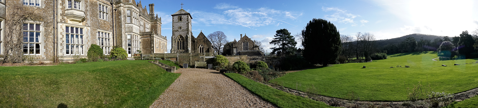
It was on the 7th of February of 2016 when 20 leading scientists from all over the world headed to the historic Wiston House in West Sussex, England, to spend four days in focused atmosphere discussing new insights in cardiovascular research: the workshop for Transdifferentiation and Tissue Plasticity in Cardiovascular Rejuvenation. Supported by the Company of Biologists, Brian Black and Jim Martin brought together experts of the field of heart development, regeneration and tissue engineering with the aim to discuss new approaches and recent findings to improve cardiac repair. In addition to the 20 senior investigators, also 10 early-stage scientists (PhD students, postdocs, and junior PIs) were selected to participate. For me in particular, with my PhD recently completed, joining this event was a great honour and a unique opportunity. I excitedly anticipated the four intense workshop days with great interest.
With the heart being such a complex and specialized tissue, repair of cardiac muscle in human is difficult to achieve. In the workshop, we were introduced to and discussed a wide range of ongoing efforts, including genomic regulation of cardiomyocyte differentiation, environmental factors involved in cardiac regeneration, as well as the contribution of different cell types in this process. Although I cannot allude to all the talks here, all participants agreed about the outstanding scientific quality of the work presented in this meeting.
Liz Robertson opened the first session of the workshop on Sunday afternoon recapitulating the origin of cardiac progenitors during embryogenesis and explaining the involvement of Eomesodermin in cardiac mesoderm specification in the mouse embryo (Costello et al., 2011). To understand myocardial differentiation, it is fundamental to define transcription factors and epigenetic modifications that specify cardiac lineages. Benoit Bruneau introduced his lab’s latest study on the coordination of three cardiac transcription factors (Nkx2.5, Tbx5 and Gata4) in the regulation of cardiac gene expression and differentiation (Luna-Zurita et al., 2016). Laurent Dupays described the interaction of the two transcription factors Meis and Nkx2.5 on a specific enhancer sequence (Dupays et al., 2015). Moreover, Brian Black explained new findings from his group about the Mef2c transcriptional regulation machinery in cardiomyocytes.
We also learned about specific approaches to elucidate the functions of distinct cellular factors active in cardiomyocytes. Guo Huang is currently investigating fetal cardiac genes, which reactivate cell cycle re-entry of adult heart muscle cells for potential regenerative repair after myocardial infarction. Jim Martin reported the implication of the Hippo pathway in cytoskeletal remodelling of cardiomyocytes in the injured heart (Morikawa et al., 2015). Finally, Kathy Ivey introduced how to study human iPSC-derived cardiomyocytes to better understand protein signalling and interaction networks as well DNA-occupancy in cellular differentiation.
Another focus during this workshop was the understanding of environmental factors, which impact cardiomyocyte behaviour and fate during cardiac repair. We learned from Eldad Tzahor how the stiffness of the extracellular matrix affects the differentiation state of cardiomyocytes (Yahalom-Ronen et al., 2015). Ahmed Mahmoud described the implication of cardiac innervation and Neuregulin signalling in the regulation of cardiomyocyte proliferation in the regenerating neonatal mouse and zebrafish heart (Mahmoud et al., 2015). The fact that many different cell types are crucial for cardiac regeneration was demonstrated by Paul Riley and Nadia Rosenthal. Nadia nicely illustrated the cellular composition of the heart and discussed recent work that demonstrates the fundamental role of macrophages during cardiac repair and regeneration (Pinto et al., 2016), while Paul Riley explained the different origins and the development of lymphatic vessels in the heart and described how this developmental program is reactivated after myocardial infarction (Klotz et al., 2015). Another approach was described by Enzo Porrello, who is seeking to understand the differences between cardiac cells at different stages of life, to unveil the mechanisms that impede adult cardiac regeneration.
Multiple talks presented studies using the zebrafish, an important model of cardiac development and regeneration due to its remarkable regenerative capacity and its transparency during embryogenesis. Didier Stainier illustrated how cardiomyocytes delaminate from the compact layer to form trabeculated myocardium in the zebrafish embryo (Staudt et al., 2014). Karina Yaniv showed beautiful movies displaying the origin of lymphatic vessels during development (Nicenboim et al., 2015). Christian Mosimann presented live imaging that traces back the cardiovascular lineages to the lateral plate mesoderm (Mosimann et al., 2015) and explained what we could learn about cardiac diseases by modelling human patient mutations in zebrafish. Ken Poss, whose lab is interested in the mechanisms of heart and fin regeneration, guided us through his journey in searching for regenerative cellular programs in the zebrafish (Kang et al., 2016). Further, Nadia Mercader spoke about distinct populations of cardiomyocytes in the early and the adult zebrafish heart and injury studies to decipher the mechanisms of myocardial regeneration.
Another key topic discussed in this workshop was how we can investigate the underlying causes of human cardiac diseases in more depth. Alessandra Moretti showed one example of how her lab studies the cause of Arrhythmogenic right ventricular dysplasia using patient-derived iPSCs. Deepak Srivastava further reported new findings about the molecular consequences of human GATA4 mutations obtained by studying iPSCs-derived cardiomyocytes from patients. An impressive finding was reported by Eric Olson: he explained how his lab achieved the repair of mutations in the Dystrophin gene, the cause of Duchenne muscular dystrophy, by CRISPR/Cas 9 technology in mice in vivo (Long et al., 2016).
I was personally very intrigued to hear about the work of the bio-engineers participating in this workshop. Nenac Bursac illustrated how his lab gains insights into cardiomyocyte functions by using in vivo assays of cardiomyocyte patches. Moreover, from Molly Stevens we learned about the versatile use of nano needles, which can deliver substances to cells or even make measurements (Chiappini et al., 2015).
In my opinion, and I am sure all participants would agree, this workshop was a tremendous success. Fascinating data, of high scientific value were presented and openly discussed. For me, this workshop was a unique experience; I met experts in the field of cardiovascular research and learned about a vast range of experimental approaches. Moreover I had the opportunity to present and discuss our latest results on the endocardial dynamics in zebrafish heart regeneration. Finally the guided tour through the amazing 16th century historical Wiston House, the windy walk in the beautiful Sussex countryside, and the experience of watching the Super Bowl for the first time, completed the great experience of the Transdifferentiation and Tissue Plasticity in Cardiovascular Rejuvenation workshop.
Here is a short video put together by the Company of Biologists on this workshop:
I am grateful to Christian Mosimann for comments on the text.
References
Chiappini, C., De Rosa, E., Martinez, J. O., Liu, X., Steele, J., Stevens, M. M. and Tasciotti, E. (2015). Biodegradable silicon nanoneedles delivering nucleic acids intracellularly induce localized in vivo neovascularization. Nature materials 14, 532-539.
Costello, I., Pimeisl, I. M., Drager, S., Bikoff, E. K., Robertson, E. J. and Arnold, S. J. (2011). The T-box transcription factor Eomesodermin acts upstream of Mesp1 to specify cardiac mesoderm during mouse gastrulation. Nature cell biology 13, 1084-1091.
Dupays, L., Shang, C., Wilson, R., Kotecha, S., Wood, S., Towers, N. and Mohun, T. (2015). Sequential Binding of MEIS1 and NKX2-5 on the Popdc2 Gene: A Mechanism for Spatiotemporal Regulation of Enhancers during Cardiogenesis. Cell reports 13, 183-195.
Kang, J., Hu, J., Karra, R., Dickson, A. L., Tornini, V. A., Nachtrab, G., Gemberling, M., Goldman, J. A., Black, B. L. and Poss, K. D. (2016). Modulation of tissue repair by regeneration enhancer elements. Nature.
Klotz, L., Norman, S., Vieira, J. M., Masters, M., Rohling, M., Dube, K. N., Bollini, S., Matsuzaki, F., Carr, C. A. and Riley, P. R. (2015). Cardiac lymphatics are heterogeneous in origin and respond to injury. Nature 522, 62-67.
Long, C., Amoasii, L., Mireault, A. A., McAnally, J. R., Li, H., Sanchez-Ortiz, E., Bhattacharyya, S., Shelton, J. M., Bassel-Duby, R. and Olson, E. N. (2016). Postnatal genome editing partially restores dystrophin expression in a mouse model of muscular dystrophy. Science 351, 400-403.
Luna-Zurita, L., Stirnimann, C. U., Glatt, S., Kaynak, B. L., Thomas, S., Baudin, F., Samee, M. A., He, D., Small, E. M., Mileikovsky, M. et al. (2016). Complex Interdependence Regulates Heterotypic Transcription Factor Distribution and Coordinates Cardiogenesis. Cell 164, 999-1014.
Mahmoud, A. I., O’Meara, C. C., Gemberling, M., Zhao, L., Bryant, D. M., Zheng, R., Gannon, J. B., Cai, L., Choi, W. Y., Egnaczyk, G. F. et al. (2015). Nerves Regulate Cardiomyocyte Proliferation and Heart Regeneration. Developmental cell 34, 387-399.
Morikawa, Y., Zhang, M., Heallen, T., Leach, J., Tao, G., Xiao, Y., Bai, Y., Li, W., Willerson, J. T. and Martin, J. F. (2015). Actin cytoskeletal remodeling with protrusion formation is essential for heart regeneration in Hippo-deficient mice. Science signaling 8, ra41.
Mosimann, C., Panakova, D., Werdich, A. A., Musso, G., Burger, A., Lawson, K. L., Carr, L. A., Nevis, K. R., Sabeh, M. K., Zhou, Y. et al. (2015). Chamber identity programs drive early functional partitioning of the heart. Nature communications 6, 8146.
Nicenboim, J., Malkinson, G., Lupo, T., Asaf, L., Sela, Y., Mayseless, O., Gibbs-Bar, L., Senderovich, N., Hashimshony, T., Shin, M. et al. (2015). Lymphatic vessels arise from specialized angioblasts within a venous niche. Nature 522, 56-61.
Pinto, A. R., Ilinykh, A., Ivey, M. J., Kuwabara, J. T., D’Antoni, M. L., Debuque, R., Chandran, A., Wang, L., Arora, K., Rosenthal, N. A. et al. (2016). Revisiting Cardiac Cellular Composition. Circulation research 118, 400-409.
Staudt, D. W., Liu, J., Thorn, K. S., Stuurman, N., Liebling, M. and Stainier, D. Y. (2014). High-resolution imaging of cardiomyocyte behavior reveals two distinct steps in ventricular trabeculation. Development 141, 585-593.
Yahalom-Ronen, Y., Rajchman, D., Sarig, R., Geiger, B. and Tzahor, E. (2015). Reduced matrix rigidity promotes neonatal cardiomyocyte dedifferentiation, proliferation and clonal expansion. eLife 4.
 (3 votes)
Get involved
Create an account or log in to post your story on the Node.
Sign up for emails
Subscribe to our mailing lists.