3rd Crick Beddington Symposium: Somites get lost without Hox keeping time
Posted by Alex Neaverson, on 7 March 2025
The 3rd Crick-Beddington Symposium, in memory of Rosa Beddington FRS (1956-2001), took place on 10th-11th February at the Francis Crick Institute, London. Rather than providing a broad summary of the event, I decided to embody the ‘Node correspondent’ persona and approach poster presenters to interview them about their research.
The symposium was very well attended, and as a result the posters were distributed across two different areas. On the lunch break of the first day, I made my way over to the quieter poster area, hoping to find a scientist willing to take part in a recorded conversation without too much background noise. Alas, enthusiasm for science was all around in the form of loud, animated discussion, which made my mission challenging!
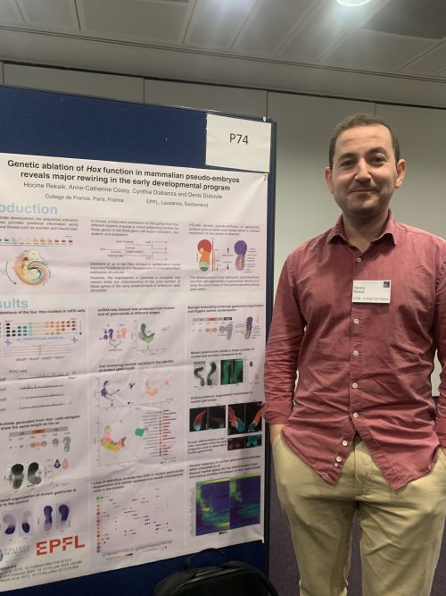
The first poster to catch my eye was presented by Dr Hocine Rekaik, who was luckily more than happy to take part. Hocine is a postdoc in the lab of Denis Duboule at College de France, Paris. The lab is interested in the function of Hox genes in vertebrate body axis development, of which the sequential activation provides the axial and paraxial tissues with positional information along the anteroposterior axis. Hox genes exhibit temporal and spatial collinearity, which means that for each gene, the timing of expression and the anteroposterior expression domain is linked to its location on the chromosome. This sequential activation, often referred to as the Hox timer, has been extensively studied in vertebrate model systems. However, the precise mechanisms linking gene expression onset with axis elongation remain elusive.
Mice, along with humans and chickens, possess four Hox gene clusters. Hocine explained that, due to the high degree of redundancy, the ideal experiment would involve deleting all of them, yet this is not possible in the mouse. Instead, they turn to the gastruloid, an embryonic stem cell (ESC)-derived model that recapitulates many aspects of gastrulation and axis elongation. Hocine explained, “These gastruloids, when they elongate, they implement the collinear expression of Hox genes, so these are really nice models to study their function and temporal expression”, which is mirrored in the gastruloid as in the embryo. Conveniently, the mouse ESCs used to create them can be modified beforehand to create a mutant line that lacks all four Hox clusters – the Hox-less clone.
Surprisingly, the Hox-less gastruloids elongate and exhibit the same anteroposterior patterning as normal, so to delve deeper into the differences between mutant and wild-type, Hocine and his colleagues performed a single cell RNA-Seq experiment. They found that at 96h and 120h of development, the Hox-less gastruloids were lacking two cell types: definitive endoderm and pharyngeal mesoderm, both of which arise from the anterior primitive streak. Yet the expression of anterior primitive streak genes was unaffected, suggesting that the streak forms as normal but its anterior derivatives are dependent on Hox expression. In accordance with this, CER1 – a crucial gene for anterior development – was significantly downregulated in the mutants. CER1 is expressed in the anterior endoderm, but also as a characteristic stripe in the newly-formed somites.
While gastruloids do express somite marker genes, they don’t exhibit the segmentation that is characteristic of somitogenesis – unless they are placed in Matrigel. “Normal gastruloids have this smooth elongation, but in Matrigel, they start to form this segmented structure” he described, and later went on to explain that this is because the Matrigel provides an extracellular matrix, which allows the cells to polarise, causing the somites to condense and epithelialise. I was surprised to learn that gene expression with and without Matrigel is the same, but Matrigel drastically changes the morphology. Hocine found that the Hox-less gastruloids tended to have fewer somites than wild-type controls, because most of them would form temporarily, then disaggregate. This was accompanied by extrusions developing at the posterior end. A pseudo-time analysis showed that the posterior somite-forming cells – derived from neuromesodermal progenitors (NMPs) – didn’t pass through all the usual cell states on their way to becoming somites, leading to the development of posterior extrusions. Hocine puts this down to the absence of the Hox clock, suggesting “there is no control over the differentiation process, so the cells start to differentiate directly – there is no gatekeeper”. However, no markers of mature, epithelialised somites were ever found in these Hox-less gastruloids.
One explanation Hocine proposed relies on the observation in the embryo that the first, most anterior somites do not give rise to segmented structures and instead, contribute to the muscles of the head, rather than the vertebrae. This region corresponds to the most anterior limit of Hox gene expression, explaining why the anterior somitic tissue is produced as normal – through disaggregation, it is simply undergoing its natural lifecycle. On the other hand, the more posterior, NMP-derived somitic tissues in altered gastruloids may have an altered trajectory and do not develop into the trunk-like mature somites seen in the control.
What’s next for Hocine’s research? He stressed that there is more work to do to understand the changes in gene expression brought about by the absence of the Hox timer. But he is excited for future experiments involving the Hox-less cell line and knows it will be very useful for the lab. They plan to do further experiments to find out how other tissues are affected, especially those involved in axial elongation, like the neural tube.
You can read Hocine’s latest article here:
Rekaik, H. et al. (2023) ‘Sequential and directional insulation by conserved CTCF sites underlies the Hox timer in stembryos’, Nature Genetics, 55(7), pp. 1164–1175. Available at: https://doi.org/10.1038/s41588-023-01426-7.
Stay tuned for more poster interviews coming soon!
 (No Ratings Yet)
Get involved
Create an account or log in to post your story on the Node.
Sign up for emails
Subscribe to our mailing lists.