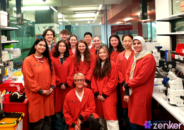
Lab meeting with the Zenker Lab
Posted by the Node, on 3 April 2025
This is part of the ‘Lab meeting’ series featuring developmental and stem cell biology labs around the world.
Where is the lab?
Jennifer: You can find the Zenker Lab at the Australian Regenerative Medicine Institute (ARMI) at Monash University. We are super lucky to call Australia, to be exact Melbourne, our home. More information is available on our Zenker lab webpage or follow us on X (formerly Twitter) @LabZenker or on LinkedIn @JennyZenker.

Research summary
The transformation of an early mammalian embryo from a tiny soccer ball-like structure into a newborn with four limbs, a beating heart, and big bright eyes is one of the most remarkable and fundamental processes of life. Errors in these early steps in development can profoundly shape a person’s lifelong ability to carry out essential tasks for everyday living. Improving the efficiency of in-vitro fertilisation (IVF) treatments 1, 2, which are expensive, physically demanding and emotionally challenging, could set up parents-to-be for the best chance of a successful cycle when it is right for them. Our research vision is to implement a new cell biological paradigm for reproductive and regenerative medicine by uncovering the real-time movements inside single cells of the living embryo and human induced pluripotent stem cells (hiPSCs).
Inside the soccer ball-like embryo resides a handful of “all-rounder” cells, known as pluripotent cells, which can give rise to any type of cell in the adult body. Using 4D (3D plus time) live-cell imaging, our research is the first in the world to conclusively demonstrate that pluripotent cells have a highly organised internal road map composed of the microtubule cytoskeleton. This road map guides the asymmetric transport of RNAs and organelles in single cells of the living mammalian embryo as a decision-making process of their future fate, becoming either part of the embryo proper or extraembryonic tissues, for instance placenta 3, 4, 5, 6, 7. Together with national and international collaborators, we have been developing innovative tools and technologies. We generated the first in vitro model of human embryos, termed iBlastoids 8, producing the most spectacular images about this new Frontier in human stem cell biology, which will continue to be a world-leading approach for many years to come 9. Furthermore, we have been pioneering the application of innovative light-switchable regulators in living 3D physiological systems to allow the spatiotemporal manipulation of the microtubule network to control the potency and function of pluripotent cells non-invasively 3, 10.
Can you give us a lab roll call?
Jennifer: I will have to start here with Louise, who is our research assistant and keeps the lab running smoothly and always stocked up. Every other lab member has their own research project. I like to design them so that team members can work easily together. There is a group of people working on the living mouse embryo. Postdoc Hongbin and two undergraduate students, Tia and Ava, are unravelling how the localisation of RNA subtypes inside single cells of the embryo contributes to the development of a healthy embryo. The project of PhD student Manlin is closely linked to it, but instead of looking at RNA subtypes, she is looking at various organelles. PostDoc Jess and undergraduate student Aya are developing new tools to interfere with the transport of RNAs or organelles by manipulating the microtubule cytoskeleton with spatial and temporal precision. PhD student Tracey and undergraduate student Zhi Bie aim to understand how similar (or not) such processes are between mouse and human model systems. Then we have PhD student Oliver, graduate student Adam and undergraduate student Micaela who are all working with human induced pluripotent stem cells (hiPSCs) to translate our discoveries in reproductive biology to regenerative medicine. This area is now even more strengthened by our newest member PhD student Minoo who is working with both systems to understand how one cell can have the capacity to give rise to all cell types an adult body is made of.
Favourite technique, and why?
Jennifer: There is only one answer — live imaging. Being able to see in real-time how a new life starts developing is simply mind-blowing, better than any science-fiction movie or so you can watch at the movies.
Apart from your own research, what are you most excited about in developmental and stem cell biology?
Jennifer: Developmental and stem cell biology is the foundation for regenerative medicine, which is one of the newest fields of research. It is an amazing time to be involved in such a rapidly evolving field. it has an exceptional potential to help the wider society worldwide by improving our capabilities to more effectively treat a wide range of diseases. I might be bias but that’s the future.
How do you approach managing your group and all the different tasks required in your job?
Jennifer: There is one quote that says it all: “Fail to prepare, prepare to fail”. It is a lot indeed, but I love to plan and prepare, being on an exact time schedule and writing to-do lists sorted by priority and time requirements. But all this only works if there is an open, clear and effective communication across all lab members.
What is the best thing about where you work?
Oliver: The best thing about where I work is being part of a great team that works on interesting and important research. We are all working together to produce cutting-edge discoveries and there is a huge amount of satisfaction that comes from that. I am also lucky that working here gives me access to high-quality facilities and instruments, as well as interactions with exceptional colleagues.
Zhi Bie: For me, it’s the opportunity to attend various internal and external seminars hosted by ARMI. Although all the research groups belong to the same institution and share a general goal in regenerative medicine, I still have a limited understanding of areas outside my own group. The weekly institute seminars provide a welcoming, knowledgeable, and meaningful platform where staff and students can engage, share ideas, and gain novel perspectives from researchers with similar backgrounds.
Hongbin: I enjoy the active exchange of scientific ideas and findings here at ARMI, through weekly internal seminars, as well as regular external seminars that invite researchers from other universities or even other countries. These seminars not only provide insights into their scientific work but also offer opportunities to discuss career paths and professional development.
Adam: In my opinion, one of the best things about working at ARMI is the people I work with. Not only are the people friendly and enthusiastic to help you however they can, but I think it is really beneficial to share a workspace that is rich in diversity because you gain valuable insight into various cultural heritages and research backgrounds.
Tia: The community, both in the institute and in the lab, is extremely special. Specifically, I like how the community continuously views science in an almost child-like wonder whilst trying to do the best we can to discover new ways to help people. Furthermore, outside of the laboratory and seminars, there is a strong sense of support that extends past just science.
Micaela: The best thing about where we work is the community among the different labs, and the always smiling faces in the tea room.
What’s there to do outside of the lab?
Louise: I started playing badminton again when I joined the lab, encouraged by my supervisor Jenny Zenker with her attitude to sport and exercise. Now I am putting more effort into this sport outside the lab with more training and I am feeling much better physically and mentally. I think that is what Jenny wants to deliver via her non-scientific role outside the lab —triathlete.
Manlin: Melbourne is one of the best student cities in the world (QS World University Rankings). As the cultural and educational hub of Australia, it offers a vibrant academic environment, diverse communities, and a high quality of life. I truly enjoy studying and living here!
Zhi Bie: Melbourne is surrounded by the sea, and I love driving along the coastal roads at sunset. Chasing the light as it fades over the ocean gives me a sense of peace and clarity—it’s windy, cozy, and feels incredibly freeing. I’m also passionate about playing squash and badminton. The time I spend on the court brings a deep sense of focus and an exhilarating sensation of explosive muscle power.
Hongbin: Outside of the lab, I enjoy Melbourne’s beautiful mountains and beaches, which help me relax, recharge, and gather fresh energy for the work ahead.
Adam: As someone who has recently joined the lab from the UK, I enjoy spending my time outside of work indulging myself in everything Australia has to offer. Whether this be early morning runs along the Yarra River trying to spot wild kangaroos, road trips to explore the Great Ocean Road and stunning rainforest or simply lazy days at the beach enjoying the Melbourne sun — no two weekends are the same!
Tia: On site, the campus is filled with many spots to explore nature while you eat lunch that are truly stunning and relaxing. Off-site, there are some amazing beaches just a short drive from the lab that are great for decompressing after a day of running experiments.
Micaela: Outside of work there are beautiful coastlines to explore where you can go swimming, surfing or hiking. At the right time of the year you can even watch humpback whales migrating up the southeast coast of Australia, which is one of the most extraordinary things to witness.
References
[1] Jin H, Han Y, Zenker J. Cellular mechanisms of monozygotic twinning: clues from assisted reproduction. Hum Reprod Update. 2024;30(6):692-705. doi:10.1093/humupd/dmae022
[2] Jin H, Zenker J (2024) Seeing is believing: Visualising the Formation of Monozygotic Twins in IVF. J Reprod Med Gynecol Obstet 9: 180.
[3] Zenker J, White MD, Templin RM, et al. A microtubule-organizing center directing intracellular transport in the early mouse embryo. Science. 2017;357(6354):925-928. doi:10.1126/science.aam9335
[4] Zenker J, White MD, Gasnier M, et al. Expanding Actin Rings Zipper the Mouse Embryo for Blastocyst Formation. Cell. 2018;173(3):776-791.e17. doi:10.1016/j.cell.2018.02.035
[5] Hawdon A, Geoghegan ND, Mohenska M, et al. Apicobasal RNA asymmetries regulate cell fate in the early mouse embryo. Nat Commun. 2023;14(1):2909. Published 2023 May 30. doi:10.1038/s41467-023-38436-2
[6] Hawdon A, Aberkane A, Zenker J. Microtubule-dependent subcellular organisation of pluripotent cells. Development. 2021;148(20):dev199909. doi:10.1242/dev.199909
[7] Stathatos GG, Dunleavy JEM, Zenker J, O’Bryan MK. Delta and epsilon tubulin in mammalian development. Trends Cell Biol. 2021;31(9):774-787. doi:10.1016/j.tcb.2021.03.010
[8] Liu X, Tan JP, Schröder J, et al. Modelling human blastocysts by reprogramming fibroblasts into iBlastoids. Nature. 2021;591(7851):627-632. doi:10.1038/s41586-021-03372-y
[9] Palacios Martínez S, Greaney J, Zenker J. Beyond the centrosome: The mystery of microtubule organising centres across mammalian preimplantation embryos. Curr Opin Cell Biol. 2022;77:102114. doi:10.1016/j.ceb.2022.102114
[10] Greaney J, Hawdon A, Stathatos GG, Aberkane A, Zenker J. Spatiotemporal Subcellular Manipulation of the Microtubule Cytoskeleton in the Living Preimplantation Mouse Embryo using Photostatins. J Vis Exp. 2021;(177):10.3791/63290. Published 2021 Nov 30. doi:10.3791/63290


 (No Ratings Yet)