An evening at Pint of Science USA – Boston/Cambridge
Posted by Vaibhav Pai, on 21 May 2015
Pint of Science is a science outreach organization that holds an annual festival across 9 countries (UK, Ireland, France, Italy, USA, Australia, Spain, Germany and Brazil) in 6 major themes, to bring the deeper, ground-breaking questions of science and the researchers working on them in contact with public at the local watering holes. The 2015 festival was from May 18th to 20th. I was fortunate enough to be invited to talk in the Pint of Science USA – Boston/Cambridge on the theme of Brain Games: From Development to Empathy gaps. It was a fantastic and engaging evening. I had the distinct pleasure of sharing the microphone with a brilliant fellow researcher Dr. Emile Bruneau from MIT who works on using neuroscience tools to discover new ways of understanding and solving conflict resolution issues across different tense areas in the world. I was given the opportunity to go first and talked about “Coding in Biological Systems: Bioelectrical Control of Tissue Identity and Anatomy” for about 10-15 mins. The transcript of my talk can be found below in this post. This was followed by a barrage of very interesting and engaging questions from the audience. Then it was Dr. Bruneau’s turn to speak and I had the distinct pleasure of being his guinea pig for an onsite experiment/demonstrations. I thoroughly enjoyed it and I believe everyone had a blast. Many deep questions and discussions followed his talk. I had the good fortune of discussing science questions, thoughts and ideas with Dr. Bruneau and some of the audience members after the event. A wonderful effort and event organization by the Pint of Science USA – Boston/Cambridge coordinators Eleana Manousiouthakis, Daniel Whittet, and Shannon Spreen and the Pint of Science community all together in making this happen. The events’ major sponsors were elife, AHA consulting engineers, USGBC Massachusetts, BGlo and D!A.
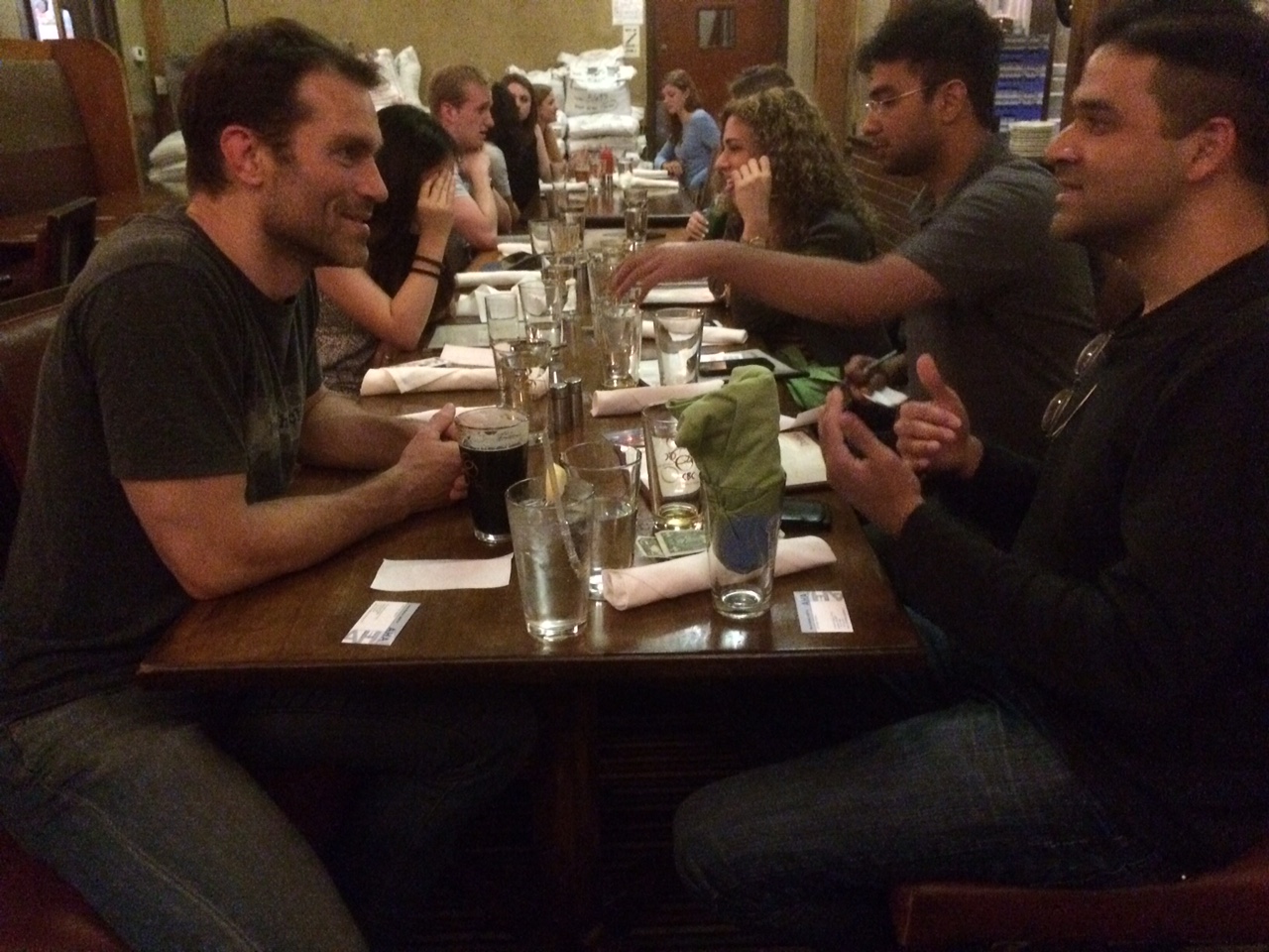
Dr.Emile and myself have a nice discussion after our talks
Title: Coding in Biological Systems: Bioelectrical Control of Tissue Identity and Anatomy
“How many of you know who a potter is? Ok for those who may not know, potter is a person who creates these beautiful pots and structures out of clay/mud. And the way that this person does this is; they have a spinning wheel on which they place a limp of moist clay and give it beautiful shape by applying forces with their hands.
Now in nature you see all kinds of beautiful shapes and structures, from that of butterflies to various birds all the way to humans. How are these shapes in nature formed? There is no external force shaping them as they develop. So where is the information for shape structure and organs stored? All animals develop from an embryo. An embryo is a single cell with one piece of genetic information. So is the information for shape and structure stored in the genetics? To find out lets do a thought experiment. Say an alien comes down to earth and hands us humans a piece of genetic information of an animal that we have never seen before. Will we be able to predict this animal’s shape, structure, organs and functions of those organs based only on the genetics of it? The answer is no! This does not mean genetics is not important. Genetics does carry important information but not all the information, only a fraction of this information. Where else then could the information for shape and structure be present? Every cell, including the developing embryo sits in a multidimensional environment where it is sensing or gathering information from all these dimensions. One of these dimensions is genetics, others including mechanical forces, chemical signals and biophysical or Bioelectric signals. We work on understanding what information is stored in bioelectrical signals and how powerful this information is.
What are bioelectrical signals? They are not externally applied electrical currents, but are endogenous (from within) currents. They are also not very fast and transient currents like one sees in the neurons. They are rather very slow currents changing over very long periods of time like hours to days! Now take any cell. It has a membrane that separates its inside from its outside. In this membrane there are proteins that act as pumps and channels. These proteins use a lot of energy to shuttle charged molecules like sodium, potassium, chloride etc, in such a way that the inside of the cell is negative and the outside positive; thus converting every cell into a battery. This voltage difference across the membrane is called membrane voltage (Vmem) and for each cell it is about 10s of mV. Now if you take a sheet of cells and look at the Vmem of cells you don’t see uniform Vmem but rather beautiful patterns of Vmem. This is highly evident in developing embryos where one sees these beautiful patterns of Vmem that occur as the embryo develops. So what information do these bioelectrical patterns contain?
To study this we use frog embryos which are experimentally practical for various reasons. We have identified one of the patterns that is critically involved in eye development. What is remarkable is that if we imprint that eye pattern elsewhere of the embryo say for example on the gut, we end up forming a whole eye on the gut of the frog tadpole. Similarly we are able to form eyes on the tail and even butt of the tadpole! What is even more remarkable is that the tadpoles are able to see through these extra eyes and their brains are able to process the visual information coming from them no matter where they physically might be present! This is done using a robot which not only records the behavior of these animals but is able to teach them and test them how much they have learned. We have now done similar experiments for bioelectric control of brain tissues as well, and are able to induce brain tissues even in the tail of the tadpole.
So what is the purpose of all this? This will help us know where and how the information for shape and structure is stored within bioelectrical signals and how it is read and implemented. This is very important for purposes of regenerative medicine, where if there is any traumatic injury we can use this to regenerate the tissues or organs. Another use is once we know this information encoding we can detect birth defects very early on before they manifest and may even be able to add that information back and correct the defect. Lastly cancers can be viewed as lump of cells that have lost the information of structure, shape and function. If we can implant the right information into the tumors we might be able to have them differentiate and incorporate into normal tissues and perform normal functions. Ok at this point I will stop and take any questions you all may have for me. “
 (1 votes)
Get involved
Create an account or log in to post your story on the Node.
Sign up for emails
Subscribe to our mailing lists.