Behind the paper: Rarely seen development of a viviparous shark – emergence of the hammerhead.
Posted by Gareth Fraser, on 12 August 2024
[This post is co-authored by Gareth Fraser and Steven Byrum.]
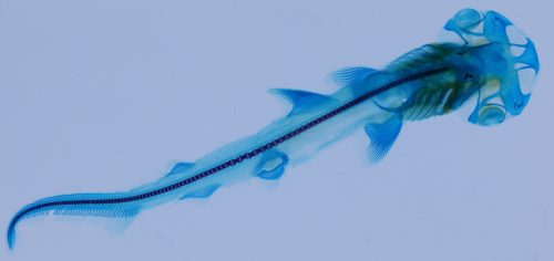
In this Developmental Dynamics paper, Steven Byrum, Gareth Fraser and colleagues present the first comprehensive embryonic staging series for the Bonnethead, a viviparous hammerhead shark. In this post, Gareth and Steven tell us more about studying these hard-to-access shark embryos and the importance of unconventional model organisms.
Why does your lab study Hammerhead shark development?
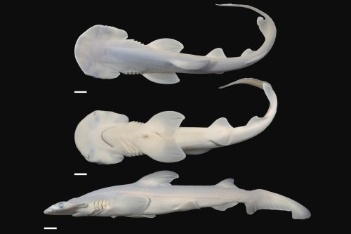
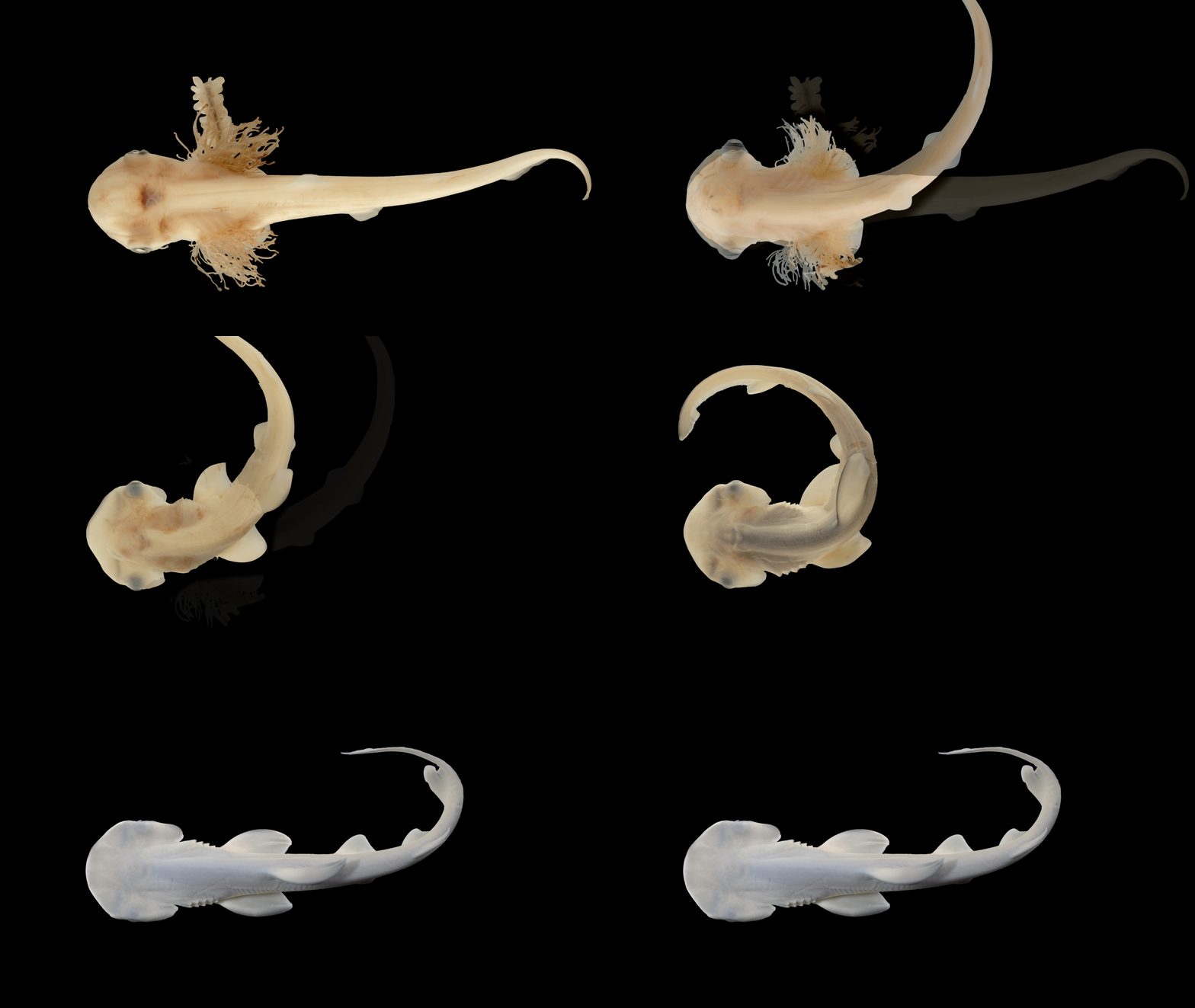
Hammerhead sharks are some of the most charismatic and enigmatic sharks in the ocean. They are also the most recognizable – owing to their characteristic hammer head or cephalofoil, a flattened and laterally expanded head with eyes present on the edges of the head “wings”. This project began with discussions within our group (Gavin Naylor, Steven Byrum and Gareth Fraser; at the Florida Museum of Natural History and the Department of Biology, University of Florida) trying to develop a project with Steven for his PhD research. Steven was interested in the development of the head in hammerhead sharks – yet no one had ever been able to study the precise stages of hammerhead embryology, only scattered observations of few embryonic stages from incidental catches (Setna and Saranghdar, 1949; Appuktan 1978) or from continued development of the established cephalofoil (Compagno, 1989). So, the project was wide-open for a more rigorous investigation. Hammerhead shark development is so difficult to study due to the inaccessible nature of their development: Hammerheads, like most sharks, give birth to live young with the entirety of their development spent in utero – a reproduction mode known as placental viviparity, where embryos obtain nutrients from their mother during embryogenesis via a placenta (somewhat similar to mammalian placental development).
How did you manage to find and study these rare shark embryos?
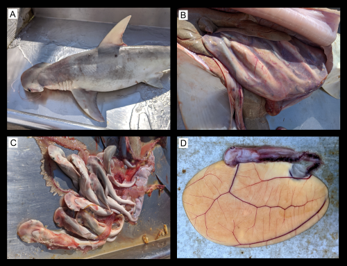
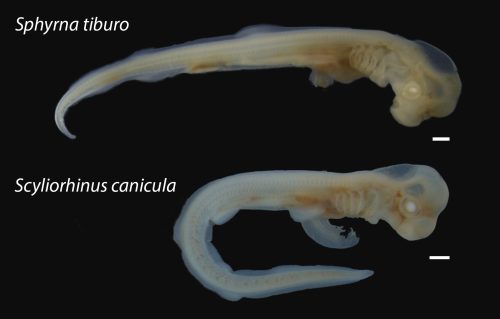
Sharks actually have three main modes of reproduction: 1) egg-laying (oviparity; with the eggs sometimes referred to as mermaid’s purses), 2) egg holding and development via yolk within the uterus (ovoviviparity or aplacental viviparity), and 3) placental viviparity. The origins and evolution of these modes of reproduction are complicated, with viviparity evolving independently multiple times from oviparous ancestors (Buddle et al, 2019; Blackburn and Hughes, 2024; Katona et al., 2023). Most of the information on shark development comes from the egg-laying varieties e.g., the well-studied Small Spotted Catshark (Scyliorhinus canicula; Ballard et al, 1993; Coolen et al, 2008; Rasch et al., 2016); egg-laying accounts for approximately 43% of all extant chondrichthyans (Compagno, 1990). Most chondrichthyan species are actually hard to study at the developmental level, being trapped inside the uterus for the entirety of embryogenesis. Therefore, access to all stages of development in viviparous hammerhead sharks is unprecedented. Until our study, the development of hammerhead sharks had mostly remained a mystery. However, we had some really wonderful collaborators, Dean Grubbs at Florida State University and Bryan Frazier at the South Carolina Department of Natural Resources, who study shark diversity at the population level and a large majority of the sharks captured are tagged and released, but a small proportion of the sharks die. These animals are retained for varied studies including diet, age and growth, reproduction, toxicology as well as developmental biology. None of the sharks from FSU or Charleston were sacrificed for this study. Occasionally, a proportion of these animals are pregnant Bonnethead shark females (Sphyrna tiburo) with embryos. The Bonnethead shark is a hammerhead shark with the smallest cephalofoil, whose populations are stable in the Gulf of Mexico and Western North Atlantic (although most hammerheads are endangered). These studies provided valuable information on the timing and seasonal periodicity of gestation, and allowed us to collect the embryos at various stages of development.
This work relied on specimen collections from wild shark populations, how difficult was this?
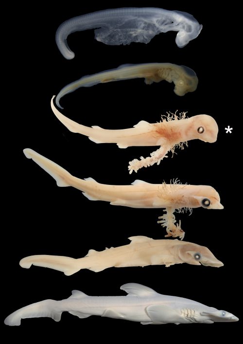
For this study, Steven collected the samples in the field, usually on board a boat with Dean or Bryan’s team with all the equipment and reagents necessary to collect and preserve the embryos. Then via CT scanning, histology and microscopy, he was able to document this rare insight into Bonnethead shark development from early head/gill arch formation to the appearance of the early cephalofoil, to the maturation of the embryo toward birth. We collected embryos over a three-year period due to the seasonal nature of Bonnethead pregnancy. Collections took place in the North Atlantic (South Carolina) and the Gulf of Mexico (Florida), within these sites, embryos begin to develop in Spring and are born in late Summer/early Fall, taking approximately 4-5 months (depending on the sea temperature and location) to complete development. However, most of this gestation period is maturation and growth, leaving a small window of opportunity each year to obtain embryos at the earlier stages. Over these three seasons (Spring 2020 – Fall 2022) we managed to collect a range of embryonic stages that included the precise time at which the hammerhead’s characteristic “hammer” starts to emerge. The one downside of relying on wild populations for specimens is there are often unsuccessful seasons, we then have to wait until the following year to obtain more embryos.
A moment in time
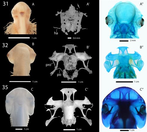
Our study identified the exact moment in development when the chondrocranium (the cartilaginous skull) starts to deviate from the ‘standard’ shark skull pattern, where we can see the early origins of the cartilaginous outgrowths that later become the characteristic hammerhead. It is this moment in developmental time that sets the Hammerhead sharks apart from all others; a moment of diversification and the origin of a developmental mutation that has ultimately allowed Hammerheads to become an incredibly successful and unique group of modern sharks.
Now that you have published the stage series for a hammerhead species, what next?
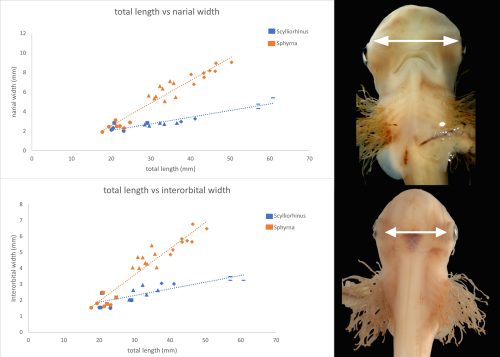
Following this unique staging series (Byrum et al, 2023) we can now push this project forward to understand the developmental genetic underpinnings of how the hammerhead forms, and why in this family of only 8 species of shark (Sphyrnidae) do their heads grow in this odd way – and importantly why no other group of sharks has evolved this strange, cephalofoil-adorned head. One of the follow up projects involves trying to understand how the developmental pathways change during this time point to allow this coordinated outgrowth of the chondrocranium in hammerheads, by comparing gene expression between Bonnetheads and Catsharks at critical moments of head development. Eventually, we can use this information to study a standard egg-laying shark that we can access embryos more readily – like the catshark – and perhaps force them genetically to form an elaborated cartilaginous skull similar to a hammerhead.
Why is it important to study unconventional model organisms?
The study of unconventional models is key to understanding the diversity of developmental processes. We should always continue to push the limits of the field and allow space to adopt unconventional models in our research programs. The wealth of untapped developmental insights in wild populations is remarkable (see a great example of this by Alexa Sadier: https://thenode.biologists.com/behind-the-paper-what-bats-can-tell-us-about-the-evolution-of-mammalian-teeth/research/), and while the standard developmental models continue to serve and have served our community well for decades, we should always look to new models that are perhaps better suited to answer particular research questions. By studying the Hammerhead shark, we can decipher the finer mechanisms of craniofacial diversity beyond what we might learn from forced mutations in the lab. Studying wild populations of unconventional models offers additional biological advantage – linking evolutionary developmental biology with true functional and ecological perspectives, made possible by the evolution of diverse morphologies. Vastly more research programs have adopted this approach in recent years, essentially “fishing” for odd or undescribed morphologies to better understand developmental diversity of animal form. Hammerhead sharks are our mutants, and this is a great example of cherry-picking natural mutants rather than making them in the lab. This is the crux of modern evo-devo – using modern techniques to study more unconventional models for the purposes of finding developmental mechanisms that drive the wonderful diversity of life on earth.
References
Appukuttan, K. K. (1978). Studies on the developmental stages of hammerhead Shark Sphyrna (eusphyrna) blochil from The Gulf of Mannar. Indian Journal of Fisheries 25, 41–52.
Ballard, W. W., Mellinger, J. and Lechenault, H. (1993). A series of normal stages for development of Scyliorhinus canicula, the lesser spotted dogfish (Chondrichthyes: Scyliorhinidae). Journal of Experimental Zoology 267, 318–336.
Blackburn, D. G. and Hughes, D. F. (2024). Phylogenetic analysis of viviparity, matrotrophy, and other reproductive patterns in chondrichthyan fishes. Biological Reviews 99, 1314–1356.
Buddle, A. L., Van Dyke, J. U., Thompson, M. B., Simpfendorfer, C. A. and Whittington, C. M. (2019). Evolution of placentotrophy: using viviparous sharks as a model to understand vertebrate placental evolution. Mar. Freshwater Res. 70, 908.
Byrum SR, Frazier BS, Grubbs RD, Naylor GJP, Fraser GJ. Embryonic development in the bonnethead (Sphyrna tiburo), a viviparous hammerhead shark. Developmental Dynamics. 2024; 253(3): 351-362.
Compagno, L. J. V. (1988). Sharks of the Order Carcharhiniformes. Princeton University Press.
Compagno, L. J. V. Alternative life-history styles of cartilaginous fishes in time and space.
Coolen, M., Menuet, A., Chassoux, D., Compagnucci, C., Henry, S., Leveque, L., Da Silva, C., Gavory, F., Samain, S., Wincker, P., et al. (2008). The Dogfish Scyliorhinus canicula: A Reference in Jawed Vertebrates. Cold Spring Harbor Protocols 2008, pdb.emo111-pdb.emo111.
Katona, G., Szabó, F., Végvári, Z., Székely, T. Jr, Liker, A., Freckleton, R. P., Vági, B., & Székely, T. (2023). Evolution of reproductive modes in sharks and rays. Journal of Evolutionary Biology, 36, 1630–1640.
Rasch, L. J., Martin, K. J., Cooper, R. L., Metscher, B., Underwood, C. J., and Fraser, G. J. (2016). An ancient dental gene set governs development and continuous regeneration of teeth in sharks. Developmental Biology. 415: 2.
Setna, S. and Sarangdhar, P. (1949). Studies on the Development of Some Bombay Elasmobranchs. Records of the Indian Mueum 47, 203–216.


 (1 votes)