Behind the paper story: From a tiny primordium to a fully developed gynoecium
Posted by Andrea Gomez, on 5 July 2024
In this post, I invite you to join me on the journey through our recent article titled “Two Orthogonal Differentiation Gradients Locally Coordinate Fruit Morphogenesis.” This story started when I joined the lab of Daniel Kierzkowski at the Institut de Recherche en Biologie Végétale (IRBV) at the Université de Montréal as a visiting PhD student in January 2020. My main goal was to learn how to perform live imaging in plants and analyze the output data. Daniel invited me to contribute to his project related to fruit morphogenesis, and I believe it fit perfectly my interest in fruit development. The central question of this project was straightforward: what are the growth patterns underlying gynoecium development from its initiation to the final shape? I started working on that project just one week prior to the COVID outbreak in Canada. I had my first confocal experiment running the day before the University of Montreal recommended all foreign internship students to go back home due to upcoming pandemics. I decided to stay and found myself “stuck” in Montreal. This situation led to my most productive period when I spent several hours, days and weeks in the microscopy room. It was just me and the confocal microscope in the entire building (a quiet solitary experience). Walking through the deserted corridors evoked a mix of sadness and loneliness, but there was also a rewarding feeling of the “perfect samples” I just imaged with the confocal microscope. We were all astonished to see that my samples could grow for two consecutive weeks, from small primordium to fully developed gynoecium (future fruit)!
Simultaneously, I juggled the demands of writing my PhD thesis, preparing one of my papers derived from my PhD, and gearing up for my thesis defense, which, due to the outbreak, had to be conducted online. It was a challenging time, but one that ultimately shaped me and my research journey in unexpected ways.
After successfully obtaining high-quality images of the gynoecium, we started the image analysis using MorphoGraphX. This part would not have been possible without the help of my colleague Elvis Branchini, whose dedication helped us segment and quantify growth in thousands of cells (from around 50 to 11000 per sample). This initial analysis represents, to our knowledge, the first comprehensive growth analysis spanning organ initiation to full expansion in plants. I take great pride in this significant achievement.
We started to dissect in detail different analyses on our gynoecium data including cell expansion, size, anisotropy, proliferation, and differentiation from each single cell. By deconstructing growth directions, both medial-lateral and longitudinal, we observed a medial-lateral growth gradient early in development. This observation contrasts with other plant organs, such as leaves, sepals, and stamen, which typically exhibit a basipetal (top-to-bottom) gradient of growth. This result was surprising for us: an organ that comes from an ancestral leaf has a different behavior. Then we started to look for explanations and formulated different hypotheses. Among these, the role of auxin — a favorite hormone in the plant scientific community — was promising within the context of our study.
Perspective from this Study:
From this study, we found that two distinct, time-shifted, and competing differentiation gradients govern gynoecium morphogenesis: an early mediolateral growth gradient and a late longitudinal growth gradient. A compelling next step would be to explore how these gradients interact. It would be fascinating to examine whether the early differentiation of the valve restricts the typical basipetal gradients from spreading through the organ, similar to what is observed in leaves, sepals, and petals. Additionally, investigating how the timing of these gradient establishments affects the final fruit shape in Brassicaceae, which exhibits a wide variety of fruit shapes, could provide valuable insights.
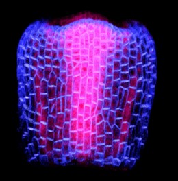
showing PIN-FORMED1 (PIN1), expression. PIN1 is
a protein carrier and a key mediator in the transport of
the plant hormone auxin. In the image, blue indicates
PIN1 expression in the epidermal layer, while pink
shows PIN1 expression in the internal layers.
My contribution:
The methods and approaches I developed for this paper are now being used in Daniel’s lab. These techniques will facilitate a more detailed investigation into fruit development across different species, improving our understanding of how fruits develop in the Brassicaceae family. This study offers a thorough and detailed atlas of growth patterns during gynoecium development. While many fascinating questions about fruit shape remain, this research paves the way for a deeper exploration of fruit development, particularly focusing on the shape and mechanical interactions within its different tissues.
My eureka moment:
Each step of the project felt like a eureka moment to me, but one has stayed with me: when we observed a full series growing continuously for two consecutive weeks. It was a delightful surprise. Additionally, each session of live imaging proved to be both gratifying and occasionally frustrating, yet undeniably worthwhile. My colleague and friend Binghan can attest to this, having shared the excitement, and participated in insightful discussions about our findings.
Bumps along the way:
Like many academic research projects, our journey with the gynoecium project was marked by challenges. We faced setbacks, moments of being stuck, and occasional frustration. I lost count of the numerous samples that did not survive or perished along the way. I recall one particular incident during my chemical treatment experiment. The plants were ready, I meticulously dissected numerous samples to maximize our chances of success. It was my fourth day of imaging, and the samples were growing really happily. Then, a disaster struck on a Sunday evening. I went to switch on the confocal microscope, and guess what? I could not initiate the system, it crashed, I wanted to cry at that moment (but I did not). Then, I lost my samples. I had to repeat the experiment again.
Similar situations occurred a couple more times, leading my colleagues to jokingly label me as having bad luck. However, I learned to cultivate resilience and approach failed experiments with a sense of humor —’Here we go, again!’— became my mantra in the realm of science.
Along the way, I learned invaluable lessons, the journey of trial and error ultimately led to new discoveries, making the effort worthwhile. When I look back and see everything we have made, I think it was all worth the effort! Despite the technical obstacles, I persevered, allowing me to expand my skills in problem-solving, critical thinking, and patience.
Our experience with the review process for our article submission was surprisingly smooth, especially compared to the tales I have heard from my colleagues. I have no complaints in that regard; both the editors and the reviewers were prompt in their responses. The comments of all the reviewers helped us improve our story. Finally, our story has found a home where it can be read.
My next step:
I have found myself profoundly inspired by this project. This experience has solidified my conviction that within science, limitless opportunities await those who approach their work with love, passion, and genuine curiosity. As scientists, we are not just observers; we are creators, empowered to innovate and explore the unknown. Following this project, I am eager to continue working on plant development and plant hormones. My focus will be on synthetic biology, and I aim to learn and utilize cutting-edge techniques such as single-cell sequencing, proteomics and CRISPR-Cas. I want to combine my knowledge of plant development and synthetic biology with one of my passions: microscopy. I’m excited to see what discoveries await on my next journey!
In the end:
The journey through this paper has significantly enhanced and refined several skills crucial for my scientific career. This accomplishment is deeply indebted to the invaluable assistance of my colleagues, who dedicated countless hours to segmenting hundreds of cells. Furthermore, the support of my friends and peers has been invaluable. Finally, I want to express my gratitude to my former boss, Daniel, for his ongoing support, insightful feedback, and discussions throughout my stay in his lab.
my favorite tool, the microscope, which was used to build this story.”
I invite you to read our story: Two orthogonal differentiation gradients locally coordinate fruit morphogenesis https://www.nature.com/articles/s41467-024-47325-1
 (2 votes)
Get involved
Create an account or log in to post your story on the Node.
Sign up for emails
Subscribe to our mailing lists.