A day in the life of an embryonic stem cell lab!
Posted by Helena Pérez Valle, on 5 February 2016
Hi everyone! I’m Helena. Some of you may know me as the current intern here at the Node, but next week, I will go back to Alfonso Martínez Arias’ lab at the University of Cambridge to continue working on my PhD. Our lab is interested in cell fate and differentiation in the context of early development. In particular, we want to understand how interactions between signals and gene regulatory networks can generate tissues and organs from single cells.
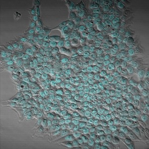
In order to study all of these phenomena, we use embryonic stem cells (ESCs). Specifically, in my project I use mouse embryonic stem cells (mESCs). mESCs are an incredibly useful system to study both cell fate decisions and early development because they are relatively easy to maintain in vitro and they are easy to access and observe; much easier than studying early development in vivo where the embryo is not always so easily accessible. Since mESCs are grown in culture, it is possible to achieve a very stringent control over the inputs the cells receive: I can control the medium the cells are grown in, the temperature, the atmosphere, what signals are applied to them and when, etc. You’d think that with so much control, I would know exactly what to expect every time I look into a tissue culture flask, but ESCs can be extremely finicky: one tiny change, for example a different batch of media or a slightly different density of cells can turn a good culture bad and throw weeks of work down the drain. Not to mention that different ESC lines all grow better in slightly different conditions, meaning each time I start using a new line, or when I culture several lines at a time, I have to adapt. However, one of the major cell drawbacks to cell culture when studying development is that the structural and mechanical constraints of the embryo are missing. Despite these limitations, mESCs are a very powerful model to study how cells respond to their environment and the types of inputs that result in maintenance of or exit from pluripotency and differentiation to specific lineages.
A typical day in an ESC lab varies considerably depending on whether one is running experiments or not, and also on the stage of the experiment. Since people in the lab are interested in how signals and gene networks interact, and because ESC culture is a model system that lends itself to producing large datasets (a single experiment can involve millions of mESCs, so potentially, millions of observations), the lab also does a lot of statistical analysis and modelling, alongside experiments. There are certain everyday tasks, however, that are fairly consistent throughout most stem cell labs, in particular tissue culture.
ESCs are fussy creatures, and require daily attention in order to maintain them in good condition in culture. This is because the function of ESCs in vivo is to divide, differentiate and build an embryo, so unless one grows them under certain conditions that promote pluripotency (i.e., with inhibitors of Wnt and FGF signalling), they are naturally inclined to differentiate. ESCs can be grown with or without feeder cells (a layer of cells underneath the ESCs that support growth, usually embryonic fibroblasts) and with our without antibiotics (to prevent infection). In our lab, we usually don’t use either: feeder cells can complicate both experiments and data analysis (for example in the case of RNA-seq experiments), and antibiotics can mask infections affecting growth and leading to contaminated experiments. ESCs are grown in flasks with cell medium, which needs to be changed every day. A lot of care has to be taken to avoid contamination: bacteria will happily grow in the nutrient-rich mESC media, eventually killing the cells. When we do have the occasional outbreak, tensions run high in the lab: a contaminated experiment is a useless one, and once a culture is contaminated, it’s not unusual for others to become infected. It also means that the tissue culture cabinets have to be meticulously cleaned with fungicide, ethanol and UV treatment – a job that no one is ever keen to take on. In our lab, we have to periodically remind each other of this, with varying degrees of severity in our reminders.
In addition to daily media changes, mESCs also need to be passaged every so often. In my case, I usually passage cells every two days, but this depends on the density of the cells in the flask. Passaging (or splitting) cells means transferring a proportion of the cells into a new flask. This is necessary in order to make sure the culture stays at the correct density, rather than becoming overgrown. This helps to maintain pluripotency and ensures that the cells are in optimal condition to perform experiments. mESCs are grown in adherent culture, meaning that the cells are attached to a matrix present on the surface of the culture flask – in our lab we use gelatin. The first thing we do when we passage cells is to apply trypsin to detach the cells from the surface of the flask. Then, we add medium to neutralise the trypsin and to dilute the cells. At this point, some people prefer to collect all the medium, spin down the cells and resuspend them before transferring into a new flask, in order to get a more accurate passage. I usually do this when I’m performing an experiment, or when I’m using a new cell line to be more accurate, but in the day to day, I will often skip this step and simply take a portion of the media I’ve used to neutralise the trypsin and transfer it to a new flask.
It is always fun to see someone new learning tissue culture. I’ve seen people with their whole heads inside a cabinet to avoid hitting the glass while working! This is obviously not the best approach for sterile technique. On one occasion, I even saw someone spray their cells with ethanol to avoid contamination. In all fairness, they achieved their goal of not contaminating their culture, although their cells died during the process! Learning tissue culture is mostly about learning good habits, although individual habits can vary from person to person. For example, in my lab, the direction pipettes are placed in the hood can be a point of contention between different people. The tissue culture cabinets can get pretty busy at times, but we all know (for the most part) when others like to do their tissue culture work, which helps with scheduling so we can all get our work done.
Once the cells have been taken care of, there are still plenty of things to do in the lab. mESC experiments usually involve growing cells in different conditions and analysing them at specific intervals. In our lab, the goal of experiments is usually to determine how the conditions used have affected the state of the cells. Has the cell exited pluripotency? If it has and it is differentiating, what is it differentiating into? How does this relate to what happens in the embryo? In order to answer these questions, we perform immunohistochemistry experiments, tracking experiments (time-lapse imaging of cell lines with fluorescent reporters for the expression of a gene of interest), flow cytometry analyses, RNA-seq, Western blots, etc. In addition, people in ESC labs spend considerable amounts of time generating new cell lines. These can be anything from cells with fluorescent reporters for the expression of genes of interest to knockout lines. As you have probably guessed, as a stem cell scientist you have to be in the lab every day when you are culturing cells. Sometimes I miss being able to go away for a weekend on a whim, but for the most part, I enjoy the flexibility. Working on the weekends can have its upsides: it means I’m not constrained by five-day experiments, and I can also use facilities that are usually busy during the week! Of course, everyone in the lab wants a weekend off once in a while, so we often “babysit” each other’s cells for a weekend. The nicer side to tissue culture is that, even though you have to do it every day, there will be days when the only thing to do is to change media – a process that can take as little as five minutes. With that done, you can spend the rest of the day catching up on reading, analysing data, imaging, and of course, on extended coffee/tea breaks chatting with your colleagues – about science of course!
 This post is part of a series on a day in the life of developmental biology labs working on different model organisms. You can read the introduction to the series here and read other posts in this series here.
 (7 votes)
One thought on “A day in the life of an embryonic stem cell lab!”
-
Pingback: Cells Weekly - February 14, 2016 - Stem Cell Assays
Leave a Reply
Get involved
Create an account or log in to post your story on the Node.
Sign up for emails
Subscribe to our mailing lists.
Read the latest Development issue
Most-read posts in May
- From comparison to mechanism: decoding heart regeneration
- A day in the life of a Reviews Editor (at Development)
- What does a Reviews Editor do?
- New evo-devo textbook ‘Eco-Evo-Devo: The Environmental Regulation of Development, Evolution, and Health’
- preLighters’ choice – A curated selection of recent preprints