Embryological Discovery in Woods Hole
Posted by Ann Grosse, on 14 July 2010
 Three weeks ago, we joined a group of twenty-four students from around the world arriving in the small town of Woods Hole, Massachusetts. We were strangers from all sorts of backgrounds, but we were drawn together by one commonality – a deep interest in developmental biology. We are all here to participate in the Marine Biological Laboratory’s Embryology course – an intensive six-week program that is in its 116th year.
The class approaches developmental biology from many perspectives using numerous model and non-model organisms. Each week is structured around a different group of animals, learning the biology and techniques which can be used to study each species, from cnidarians to flies to worms to frogs (and many more!). A typical day starts with a morning lecture presented by a leading scientist, who describes his or her research in the context of the development of the study organism. Following lecture, we have a student driven discussion. Over the years this has come to be known as the “sweat box” because of the probing nature of the questions asked by the students! In the afternoon, lab section begins, and we keep working until late into the night learning new techniques for each organism, as well as using a range of imaging techniques to visualize our results. The students also design and carry out our own research projects, which is the most exciting part of all!
Introduction to each of our research interests:
Laurel Hiebert: I am a second-year graduate student at the Oregon Institute of Marine Biology, University of Oregon, where I am studying the emergence of antero-posterior patterning in a novel larval body plan – the pilidium larva of marine ribbon worms (phylum Nemertea). I have broad interests in evolution of morphological diversity, and I am particularly excited about the opportunity to learn about development in a great variety of organisms.
Sorrel Bickley: I work at the National Institute for Medical Research in London, where I am in the first year of my Ph.D. studying the development of the limbs and the pectoral girdle. I am broadly interested in organogenesis and the processes and pathways that regulate cell differentiation and migration to form a functioning structure. In my lab I work with chick and mouse, so I am excited about the opportunity to try out new techniques in these organisms as well as learning about other species.
Ann Grosse: I am a graduate student in Deborah Gumucio’s lab at the University of Michigan. I study the morphogenetic changes responsible for remodeling the embryonic mouse intestinal epithelium to form a functional absorptive epithelial layer. More generally, my interests include answering embryological and developmental questions of cell polarity, shape, and movement using animal models, live imaging, genetic manipulation, and developmental techniques. Because I will soon be graduating, I am excited to interact and learn from faculty and students who are leaders in their respective fields. Experimentally, I look forward to the animal models most suited for live imaging: C. elegans, Xenopus, zebrafish, and sea urchin.
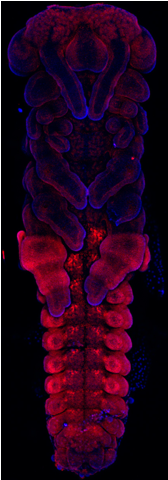
We will update this blog each week to discuss the work we have been doing on the course and also to show some of the images we have captured. As an introduction we would like to share this beautiful grasshopper embryo image, which was stained with an Ultrabithorax antibody (in red) and DAPI (in blue). We are very thankful to the Company of Biologists, who provided scholarship funds for the three of us.


 (2 votes)
Sounds very good and interesting. I hope some day I have an opportunity to join such a course, too. Company of Biologists is very supportive to graduate students. Yeah…..