Healing an injured heart
Posted by Erin M Campbell, on 5 May 2011
Regenerative medicine and stem cell research go hand-in-hand when it comes to dreaming up future strategies for treating disease and injury in humans. Today’s image is from a recent Development paper discussing how damaged heart tissue regenerates in zebrafish, and serves as a great model for devising strategies to help human heart attack patients.
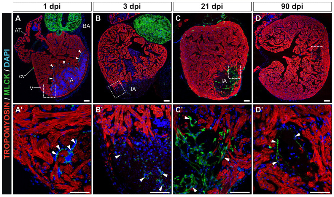
 When a person suffers a heart attack, white blood cells move into the injured area of the heart and create scar tissue. This scar tissue is important to maintain the structural integrity of the heart, but causes long-term changes in the heart’s architecture that may lead to heart failure. A recent paper in Development looks at this process in zebrafish, and describes how the zebrafish heart can undergo regeneration after injury to cardiac tissue. In this paper, researchers used cryocauterization to cause localized injury to the heart that appears similar to that seen in humans after a heart attack. Cryocauterization caused myocardial cell apoptosis within the injured area, followed by formation of scar tissue, followed by complete regeneration. This regeneration included key cardiac tissue types, including epicardium, myocardium, endocardium and coronary vasculature. This amazing regeneration ability of the zebrafish heart may provide a framework for how this process may be engineered for human patients after suffering heart attacks.
The images above show zebrafish heart tissue after injury (dpi = days post-injury), with bottom images showing higher magnification views of the boxed regions. The injured area (IA) lacks tropomyosin staining (red). Shortly after injury (A), the presence of Mlck (myosin light chain kinase, green) at the border of the injury indicates the presence of activated platelets, which promote scar formation. After a few days, the Mlck-positive cells in the injured area (B,C) indicates the presence of smooth muscle scar tissue. Many days after injury (D), the lack of Mlck suggests that the scar tissue has been replaced with new, healthy tissue.
For a more general description of this image, see my imaging blog within EuroStemCell, the European stem cell portal.
![]() Gonzalez-Rosa, J., Martin, V., Peralta, M., Torres, M., & Mercader, N. (2011). Extensive scar formation and regression during heart regeneration after cryoinjury in zebrafish Development, 138 (9), 1663-1674 DOI: 10.1242/dev.060897
Gonzalez-Rosa, J., Martin, V., Peralta, M., Torres, M., & Mercader, N. (2011). Extensive scar formation and regression during heart regeneration after cryoinjury in zebrafish Development, 138 (9), 1663-1674 DOI: 10.1242/dev.060897


 (5 votes)