In Development this week (Vol. 141, Issue 21)
Posted by Seema Grewal, on 21 October 2014
Here are the highlights from the new issue of Development:
Two top tips for angiogenesis
 The widely accepted model of angiogenic sprouting proposes that a single cell – the tip cell – is found at the leading edge of vessel sprouts. Now, Victoria Bautch and colleagues describe an alternative blood vessel topology in which multiple endothelial cells (ECs) constitute the tip of sprouting blood vessels and polarize to promote lumen formation (p. 4121). By mosaically labelling ECs in both in vitro and in vivoangiogenesis assays, the researchers first demonstrate that sprouts often contain two cells at their tips, and that both cells extend filopodia. Live imaging studies show that, as reported previously, these tip cells switch positions, but the cell-cell overlap is largely maintained throughout switching. The researchers further report that tip cells polarize along their longitudinal cell-cell border; this border is characterized by apical polarity markers and is the site of lumen formation. Finally, the authors show that the loss of an atypical protein kinase C (aPKC) isoform disrupts EC overlap and lumen formation, highlighting a role for aPKC in angiogenic sprouting. Together, these observations suggest that the paradigm of a single cell at the tip of developing blood vessels requires revision.
Fat body shapes up the trachea
 Development of the Drosophila tracheal system – the fly respiratory network – is known to require the chitin deacetylase serpentine (Serp), which is expressed in tracheal epithelial cells. Here, Shigeo Hayashi and co-workers show that Serp derived from the Drosophila fat body, which is functionally equivalent to the mammalian liver, is transported to the tracheal lumen and is required for tube morphogenesis (p. 4104). They first show that Serp is expressed in the fat body and that Serp levels accumulate in fat bodies of the rab9 and shrub vesicle trafficking mutants. By contrast, Serp levels in the tracheal lumen of these mutants are reduced, suggesting that Serp is secreted into the haemolymph and is taken up by tracheal cells. Importantly, the researchers show that fat body-derived Serp is able to rescue the tracheal defects observed in serp mutants. These studies suggest that Serp should be added to the expanding repertoire of proteins that the fat body supplies to other organs and highlight the role of the fat body in regulating the development of other organs.
Nf2: guiding axons across the midline
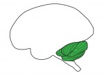
 The cerebral hemispheres are connected by the largest axonal tract in the mammalian brain: the corpus callosum (CC). During CC development, axons from one hemisphere navigate across the midline, channelled along their way by guidepost cells that secrete guidance cues, to reach specific targets on the other hemisphere. Neurofibromatosis type 2 (Nf2/Merlin) mouse mutants show a complete absence of the CC but how Nf2, a signalling protein involved in various cellular processes and signalling pathways, controls axonal pathfinding is currently unclear. Using Nf2-conditional knockout mouse models, Xinwei Cao and colleagues show that, surprisingly, Nf2 is not required in callosal neurons or their progenitors but is required in midline neural progenitors that generate guidepost cells (see p. 4182). The authors reveal that Nf2 controls guidepost development and the expression of Slit2, a major signalling cue secreted by guidepost cells, through the suppression of YAP, an effector of the Hippo pathway. These findings represent an exciting step forward in the molecular understanding of midline formation and brain wiring, as well as uncovering an intriguing previously undescribed function for the Hippo pathway.
Getting to the heart of slow conducting cardiomyocytes
 During a heart beat, an electrical impulse leads to the contraction of the upper part of the heart: the atria. The electrical impulse slows down through the atrioventricular junction (AVJ) before resuming rapid propagation to induce contraction of the lower part of the heart: the ventricles. This contraction delay, combined with the presence of cardiac valves, is crucial for unidirectional blood flow in the heart and is altered in various heart diseases. How is this slow conducting property established and restricted to the AVJ? On p.4149, Takashi Mikawa and colleagues discover that, contrary to the current hypothesis, the AVJ does not maintain juvenile slow conduction; instead, AVJ conduction velocity is plastic and determined by its proximity to the endocardium (the inner lining of the heart). They further show that the cardiac jelly (an extracellular martix-rich deposit that accumulates during valve formation) acts as a crucial physical barrier separating the AVJ from endocardial signals that induce a fast conduction phenotype. The authors thus uncover an exciting mechanism whereby valve formation and the delay in chamber contraction are developmentally linked, and open new perspectives for understanding heart development and congenital diseases.
PLUS…
Development of the cerebellum: simple steps to make a ‘little brain’
 The cerebellum is a pre-eminent model for the study of neurogenesis and circuit assembly. In recent years, our understanding of cerebellar growth has undergone major recalibration, and insights from a variety of species have contributed to an increasingly rich picture of how this system develops. Here, Thomas Butts, Mary Green and Richard Wingate review these advances. See the Review on p. 4031
Making designer mutants in model organisms
 Recent advances in the targeted modification of eukaryotic genomes have unlocked a new era of genome engineering. From pioneering work using zinc-finger nucleases, to the advent of TALEN and CRISPR/Cas9 systems, we now possess the ability to analyze developmental processes using sophisticated genetic tools. Here, Stephen Ekker and colleagues summarize the common approaches and applications of these still-evolving tools. See the Review on p. 4042
 (1 votes)
Get involved
Create an account or log in to post your story on the Node.
Sign up for emails
Subscribe to our mailing lists.