In Development this week (Vol. 141, Issue 22)
Posted by Seema Grewal, on 4 November 2014
Here are the highlights from the current issue of Development:
Meristem maintenance is KNOX so simple
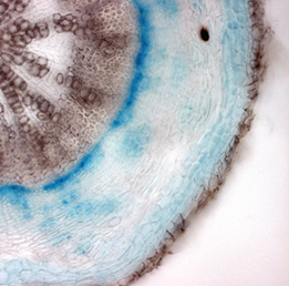 Class I KNOX transcription factors, such as SHOOT MERISTEMLESS (STM) and KNAT1, are known to play a role in the plant shoot apical meristem (SAM), where they are thought to prevent differentiation and hence promote stem cell maintenance. Now, on p. 4311, Urs Fischer and colleagues uncover a role for STM and KNAT1 in another Arabidopsis meristem – the vascular cambium, which is a lateral meristem that gives rise to xylem and phloem cells. They first show that STM and KNAT1 are expressed in undifferentiated cambial cells but also in differentiated phloem and xylem cells. The researchers further demonstrate that xylem fibre formation is reduced in stm and knat1 mutants, suggesting that STM and KNAT1 promote the differentiation of cambial derivatives. In addition, they report that STM and KNAT1 regulate xylem differentiation via transcriptional repression of BLADE-ON-PETIOLE 1 (BOP1) and BOP2, which are not expressed in the SAM. Together, these findings demonstrate that, in contrast to their function in the SAM, STM and KNAT1 promote cell differentiation in the cambium, suggesting that the exact role of these transcription factors in other meristems needs re-examination.
TBX1: at the heart of epithelial properties
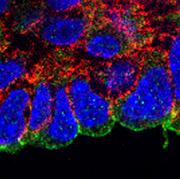 During its development, the heart tube undergoes rapid elongation, fuelled by the addition of cardiac progenitors from the second heart field (SHF). The gene regulatory networks governing SHF formation have been studied extensively, but little is known about the basic cellular features of SHF cells. Now, Robert Kelly and co-workers show that the transcription factor TBX1, which is implicated in both normal SHF development and congenital heart defects, regulates the epithelial properties of mouse SHF cells (p. 4320). Using immunofluorescence microscopy, they first show that SHF cells in the dorsal pericardial wall constitute an apicobasally polarised epithelium. Transmission and scanning electron microscopy reveal the presence of monocilia on the apical surface of SHF cells and of actin-rich filopodia on their basal surface. Using live-imaging of thick-slice cultures, the researchers demonstrate that these filopodia are dynamic, extending towards and making contact with surrounding tissues. Importantly, they report that TBX1 plays a crucial role in regulating these epithelial cell features; cell shape, cell polarity and filopodia dynamics are perturbed in Tbx1-/- mutants. These exciting findings suggest that TBX1-mediated control of epithelial state is crucial for heart development.
Mesogenin 1 masters the presomitic mesoderm
 During development, neuromesodermal (NM) stem cells give rise to both neural cells and paraxial presomitic mesoderm (PSM) cells, but what dictates PSM fate? Here, Terry Yamaguchi and colleagues show that a single transcription factor – mesogenin 1 (Msgn1) – acts as a master regulator of PSM development (p. 4285). They show that the overexpression of Msgn1 in mouse ESCs cultured as embryoid bodies (EBs) is sufficient to drive PSM differentiation. Microarray and ChIP-seq analyses of Msgn1-overexpressing EBs confirm that Msgn1 controls the expression of key regulators of PSM development, including those involved in epithelial-mesenchymal transition and segmentation. Importantly, the researchers demonstrate that Msgn1 overexpression in NM stem cells in vivo biases fate towards the PSM; the contribution of these cells to the neural tube is reduced while the number of PSM cells is dramatically increased. Finally, the authors show that Msgn1 overexpression can partly rescue the PSM differentiation defects observed in Wnt3a−/− embryos, suggesting that Msgn1 functions downstream of Wnt3a as master regulator of PSM fate. Given the role of the PSM as a precursor for a multitude of cell types, this finding has important implications for the fields of cellular reprogramming and regenerative medicine.
Basonuclin 2: a regulator of spermatogenesis
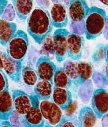 Embryonic germ cells display strikingly different fates with regard to mitosis and meiosis, depending on their sex. In female mice, germ cells switch from mitosis to meiosis shortly after reaching the foetal gonad where they generate the lifelong pool of oocytes. However, in males, meiosis and mitosis are actively repressed, and germ cells remain quiescent in the gonad until birth, when they resume mitosis and start generating spermatocytes. Here (p. 4298), Philippe Djian and colleagues demonstrate that Basonuclin 2, an extremely conserved transcription factor specifically expressed in male germ cells, suppresses meiosis. More surprisingly, they also show that Basonuclin 2 is required for mitosis repression and, later in life, for meiosis progression during spermatogenesis and maintenance of the spermatogonial stem cells that ensure spermatocyte production during life. Furthermore, Basonuclin 2 is necessary for the expression of DNMT3L, a key protein that is involved in spermatogenesis, and for the repression of meiotic genes (Stra8, Msx1 and Msx2) that are normally expressed in female germ cells. These findings, which uncover a new regulator of male gametogenesis, are likely to further our understanding of spermatogenesis in humans.
From embryonic stem cells to gastruloids: early development in a dish
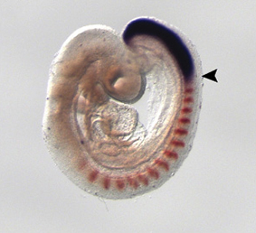
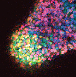 One of the first patterning events of embryogenesis occurs during gastrulation: three-dimensional (3D) cell movements reorganise the embryo, a mass of morphologically similar cells, into an axially organised structure with three germinal layers (endoderm, mesoderm and ectoderm). To date, two-dimensional (2D) culture models have failed to recapitulate such complex cell behaviours linking cell movement to cell fate. Here (p. 4231), Alfonso Martinez Arias and colleagues show that 3D aggregates of mouse embryonic stem cells cultured in mesendoderm-promoting medium undergo cell movements, axial organisation and germ layer specification, features reminiscent of gastrulation. They demonstrate that the expression of endoderm (Sox17, Fox2A) and early mesoderm (Brachyury) markers becomes polarised in these aggregates. Later, cells originating from the Brachyury-expressing ‘territory’ are extruded from the aggregate. These ‘gastruloids’ thus present a powerful tool that can be used to study early embryonic tissue specification in a dish, an unprecedented feat in vitro.
PLUS…
Reactive oxygen species and stem cells
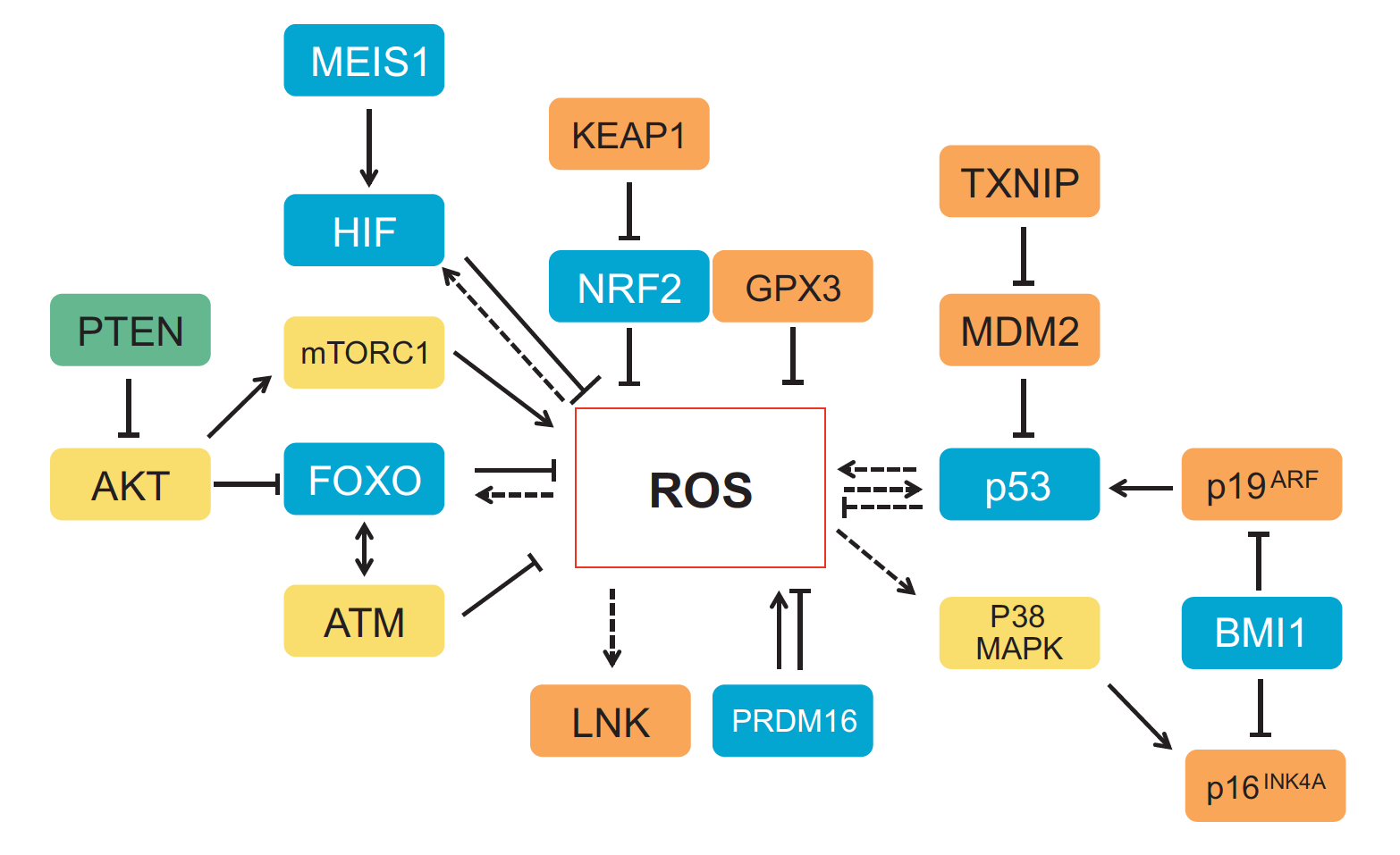 Recent work suggests that reactive oxygen species (ROS) can influence stem cell homeostasis and lineage commitment. In this Primer, Ghaffari and colleagues provide an overview of ROS signalling and its impact on stem cells, reprogramming and ageing. See the Primer on p. 4206
Chemokines in development and disease:
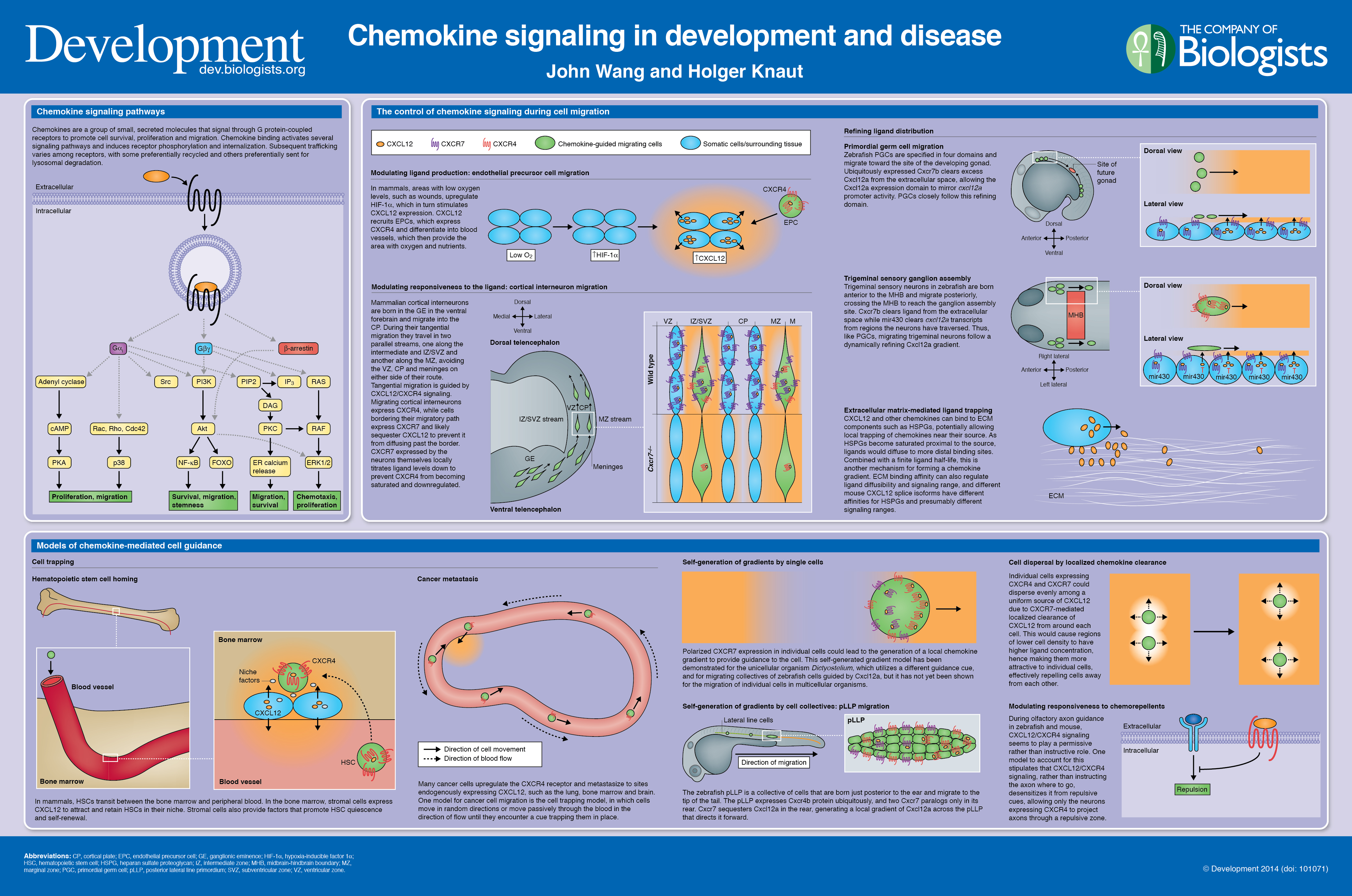 In our latest poster and companion article, Wang and Knaut provide an overview of chemokine signalling and some the chemokine-dependent strategies used to guide migrating cells. See the poster on p. 4199
Leaf development and morphogenesis
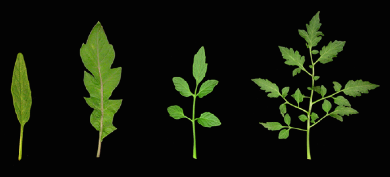 The development of plant leaves follows a common basic program, which can be modulated to generate a diverse range of leaf forms. Bar and Ori review recent work examining how plant hormones, transcription factors and tissue mechanics influence leaf development. See the Review on p. 4219
 (No Ratings Yet)
Get involved
Create an account or log in to post your story on the Node.
Sign up for emails
Subscribe to our mailing lists.
Read the latest Development issue
Most-read posts in May
- From comparison to mechanism: decoding heart regeneration
- A day in the life of a Reviews Editor (at Development)
- What does a Reviews Editor do?
- New evo-devo textbook ‘Eco-Evo-Devo: The Environmental Regulation of Development, Evolution, and Health’
- preLighters’ choice – A curated selection of recent preprints