In Development this week (Vol. 142, Issue 24)
Posted by Seema Grewal, on 15 December 2015
Here are the highlights from the current issue of Development:
New markers for human endoderm differentiation

The generation of mature cell types from pluripotent stem cells (PSCs) relies on lineage-specific markers to track and enrich for distinct cell populations. During hepatocyte differentiation, the induction of the definitive endoderm is a crucial step; however, to date there are no markers that exclusively recognise differentiating human endoderm, nor any that can select for hepatocyte-fated cells from within this population. Now, on p. 4253, Gordon Keller and colleagues report on the identification of two antibodies that can be used to identify and select for cells undergoing human endoderm differentiation from PSCs. The authors name the antibodies HDE1, which exclusively marks the entire endoderm population as it emerges, and HDE2, which marks emerging hepatic progenitors and mature hepatocytes. The authors show that the extent of HDE1 reactivity correlates with hepatic potential, and, importantly, that the two antibodies work across numerous different human PSC lines. This exciting breakthrough enables the individual monitoring of both endoderm induction and hepatic specification, leading to a more efficient protocol for directed differentiation of human hepatocytes from PSCs.
Local mechanism for decline in stem cell proliferation

As tissues age, the rate at which endogenous stem cells proliferate is known to decline, leading to prolonged periods of quiescence and fewer stem cell progeny. Nutrient status is a key regulator of stem cell proliferation; however, it remains fairly constant across all tissues in the body, whereas rates of stem cell proliferation can vary widely among tissues. In order to explain this phenomenon, there must be a mechanism for the local regulation of stem cell proliferation, but so far this has remained elusive. Now, on p. 4230, Jean-Claude Labbé and colleagues uncover a process that mediates a local decline in germline stem cell (GSC) proliferation in C. elegans. The authors show that an accumulation of differentiated progeny, in this case oocytes, causes a decrease in GSC proliferation rates. Interestingly, this induced GSC quiescence is caused by local inhibition of insulin/IGF-1 signalling mediated by DAF-18/PTEN, but not DAF-16/FOXO, signalling downstream of oocyte accumulation. Since insulin/IGF-1 signalling operates in all animals including humans, these results represent an exciting breakthrough in our understanding of how stem cell proliferation and quiescence can be regulated in a tissue-specific manner, and may have important implications for disease.
Size matters: lessons from the planarian on organ scaling

Despite many advances in understanding stem cell regulation and growth signalling, the developmental mechanisms controlling organ size attainment remain elusive. How does an organism know when the appropriate number of cells in an organ has been reached, either during development or regeneration? In this issue (p. 4217), Christian Petersen and Eric Hill describe a novel system to investigate whole cell number control during organ regeneration in the planarian. In their study, the authors report how a specific ratio of brain neurons to body size is maintained during periods of organismal growth, shrinking and regeneration. Using this foundation, the authors demonstrate how wnt11-6 (expressed at the posterior of the brain) and the Wnt inhibitor notum (expressed at the anterior of the brain) co-regulate each other and ultimately determine brain size. Further, the authors show this is not through cell proliferation or death, but through a pathway involving canonical and non-canonical Wnt signalling that influences neural progenitor numbers. Taken together, these results illustrate a genetic mechanism for the loss of cell number during regeneration and show how stem cell regulation can be responsible for organ size reduction.
MAP(K)ping out binary fate decisions in ESCs

One of the earliest events in mammalian development occurs when the inner cell mass segregates into two distinct cell populations: the epiblast (Epi) and the primitive ectoderm (PrE). Much is known regarding the molecular and signalling pathways that regulate this early fate decision, but what remains unclear is how these inputs are integrated into the molecular circuitry in order to regulate precisely the temporal and spatial emergence of these two cell lineages. Now, on p. 4205, Alfonso Martinez Arias and colleagues investigate this question using multicolour single-cell quantitative assays and mathematical modelling, and demonstrate a dual role for FGF/MAPK signalling in the decision between PrE and Epi cell fates. Firstly, the authors show that in order for GATA factors to activate the PrE gene expression programme, the FGF/MAPK pathway must be inhibited. Secondly, the authors demonstrate that MAPK signalling also sets the threshold level of GATA transcription factors required to specify the PrE lineage, and thereby controls the proportion of PrE cells. These data are used to parameterise a binary switch network model that describes the mechanism and predicts how MAPK input is incorporated into the network.
On the origins of organ-specific vessel formation

Every organ must be properly vascularised in order to receive nutrients and signals, and to remove waste. Although it is clear that some blood vessels show features specific to their organ of origin, it is not yet understood how organ-specific vessels arise during embryonic development, nor what the molecular mechanisms are that regulate their formation. In this issue (p. 4266), Karina Yaniv and colleagues use the zebrafish subintestinal plexus, a vascular bed that gives rise to the vessels of the gut, liver and pancreas, to dissect the early cellular and molecular events of organ-specific vascularisation. The authors show a common origin for all cells within the subintestinal plexus: a pool of specialised angioblasts located in the floor of the posterior cardinal vein. The authors demonstrate that these specialised angioblasts undergo two rounds of migration and differentiation, which are regulated by BMP and VEGF, respectively. Interestingly, Notch is required only during later stages of subintestinal plexus development, and not earlier. These results provide new insights into the origins of organ-specific blood vessels and showcase the zebrafish subintestinal plexus as a powerful model for characterising this phenomenon.
PLUS…
Tendon development and musculoskeletal assembly: emerging roles for the extracellular matrix
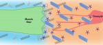 Tendons are ECM-rich structures that interconnect muscles and bones. Here, Subramanian and Schilling review how intrinsic mechanisms as well as extrinsic factors, such as mechanical force, regulate the ECM to control tendon development and maturation. See the Review on p. 4191
Featured movie
Our latest feature movie shows ESCs expressing a H2B-Cerulean transgene under the control of the CAGS promoter. Read the paper by Martinez Arias and colleagues, where they use mouse ESC lines to study the mechanism underlying the decision between the Epi and the PrE fate, on p. 4205
 (No Ratings Yet)
Get involved
Create an account or log in to post your story on the Node.
Sign up for emails
Subscribe to our mailing lists.