Interview with Arthur Boutillon: Editor’s Choice of the MBL Embryology course image competition
Posted by Andrea Murillo, on 13 October 2025
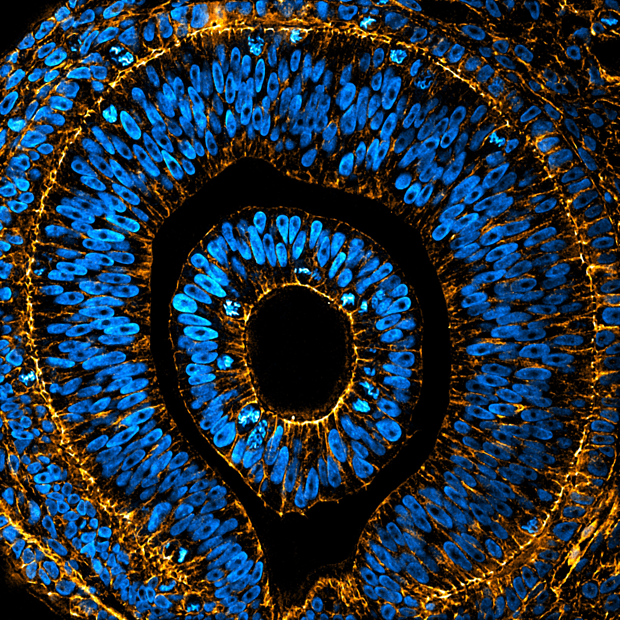
This year brought the return of our image competition with the MBL Embryology course at Woods Hole. Twenty impressive submissions were received from the 2025 class of students, with images ranging from polychaete worms to butterflies, squids and mice. Here, we interview Arthur Boutillon, the Editor’s Choice winner of the image competition with his submission, ‘Embryonic eye of Anole lizard’. As our Editor’s Choice, Arthur’s image was featured on the cover of a recent issue of Development.
Arthur Boutillon
Embryonic eye of Anole lizard stained for nuclei (DAPI, blue) and F-actin (Phalloidin, orange), imaged by spinning disc confocal microscopy and processed using ImageJ.
Can you describe your research career so far?
My research career began during my Bachelor’s studies at the École Normale Supérieure in Paris, France, where I had the opportunity to work with different model organisms through several internships, all focused on morphogenesis.
I then pursued a PhD in Nicolas David’s team at The Laboratory for Optics and Biosciences, École Polytechnique, Palaiseau, France. Working with zebrafish embryos, my favourite model organism so far, I studied the collective movement of cells during gastrulation using a combination of classical approaches (e.g., grafts) and more advanced techniques (e.g., 3D laser ablations), along with extensive live microscopy. We discovered a novel mechanism by which cells coordinate over long distances, a phenomenon we named “guidance by followers”.
Can you tell us about your current research?
After defending my PhD at the end of 2021, I moved to Dresden, Germany, to join Otger Campàs’ group in the Cluster of Excellence Physics of Life as a postdoctoral researcher. Here, I study the mechanics of morphogenesis, still using zebrafish as my model system.
By combining physical measurements, quantitative imaging, and genetic perturbations, I investigate the mechanics of somite boundary formation, work that is currently under revision for publication. I also study the link between signaling and the acquisition of tissue mechanical properties.
Attending the Embryology Course at Woods Hole broadened my perspective on the value of different model organisms. I am now developing research projects centered on mechano-evo-devo to apply for PI positions.
What is your favourite imaging technique/microscope?
Anything that allows me to watch development live!
That said, I have a soft spot for the versatility of point-scanning microscopy.
With our trusted LSM980, I can image both live and fixed samples and perform laser ablation, optogenetics, FRAP, FCS, and more, all on the same instrument!
What are you most excited about in microscopy currently/in the future?
I’m excited about all kinds of new developments: every microscopy technique has its strengths and limitations, and it’s ultimately about finding the best fit for each question. What excites me even more are the tools that help with segmentation and image analysis. I’m constantly amazed by what open-source platforms like StarDist, Cellpose, or Mastodon can achieve.
My dream is to see a unified, open-source platform that integrates these tools seamlessly, something that would save long hours of image analysis after acquisition!
 (1 votes)
Get involved
Create an account or log in to post your story on the Node.
Sign up for emails
Subscribe to our mailing lists.