Interview with Nicole Roos and Anthony Wokasch: winners of the MBL Embryology course image competition
Posted by the Node, on 10 November 2025
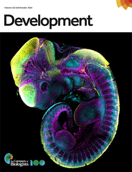
Last September marked the return of our image competition with the MBL Embryology course at Woods Hole. The 2025 cohort submitted impressive images, ranging from polychaete worms to butterflies, squid, and mice, using a range of microscopy techniques. Here, we interview Nicole Roos and Anthony Wokasch, the winners of the popular vote of the image competition with their submission, ‘Mouse embryo’. As winners of the image competition, their submission was featured on the cover of a recent issue of Development.
Can you describe your research career so far?
Nicole: As a kid I loved conducting at-home experiments, visiting the science museum and attending my chemistry and biology classes, so pursuing a career in science has always been a no brainer for me. I started my research career as a freshman at The University of Texas at Dallas, USA, while pursuing my BSc in Biochemistry. Inspired by a family member who survived breast cancer, I joined the lab of Dr Nikki Delk to study chronic inflammation in tumor microenvironments. My interest in genetics was sparked while investigating mutations that affect chromatin remodeler function at the University of Texas Southwestern Medical Center, USA, with Dr Laura Banaszynski. Now, I am a fourth year PhD candidate working with Dr Leila Rieder in the Genetics and Molecular Biology program at Emory University, USA.
Anthony: I began my research career at American University, USA, where I completed a joint BSc/MSc in Biology. During my time there, I did my Master’s thesis in the lab of Naden Krogan, where I characterized the role of the floral boundary gene SUPERMAN (SUP) in a transcriptional mechanism which regulates gene expression and floral organ patterning of the reproductive organs in the flower of Arabidopsis thaliana. Following completion of my Master’s, I became a fellow at the National Cancer Institute (NCI) in the lab of Dr Peter D. Aplan. There I tested the effects of a DNA methyltransferase (DNMT1) inhibitor in a murine model for Myelodysplastic Syndrome (MDS). However, at this point my interest in development was overwhelming and led me to pursue a PhD at Vanderbilt University, USA, where I am a PhD candidate in my fourth year.
Can you tell us about your current research?
Nicole: At Emory, I use the powerful model system Drosophila melanogaster (fruit flies) to study how histone genes, which encode proteins that package and organize DNA in the nucleus, are regulated in embryogenesis and throughout development. Histone gene regulation is uniquely regulated throughout development, and this regulation is carried out by a nuclear body called the histone locus body. Not only do I get to study genetics, but through my research I took interest in developmental biology. This compelled me to apply to the Embryology course offered by the Marine Biological Laboratory (MBL) in Woods Hole, USA, where I discovered a strong desire to study regeneration and/or evolutionary developmental biology in the future.
Anthony: My current research, under the guidance of Dr Maureen Gannon focuses on how the Pdx1 transcription factor and its C-terminal interacting factors, Oc1 and SPOP, regulate the choice between proliferation or differentiation of early pancreatic progenitor cells. This work has deepened my understanding of how epigenetic and molecular factors control cell fate decisions during organogenesis.
What is your favourite imaging technique/microscope?
Nicole: The majority of my research at Emory University requires widefield fluorescence microscopy, so I was excited to take the Embryology Course at the MBL to learn various confocal microscopy techniques. I quickly became fascinated with live imaging using various spinning disc confocal microscopes (my favorite being the Nikon Yokogawa W1 spinning disc) and collaborated with other students to live image processes such as sea urchin egg and sea star sperm cross-fertilization (sea sturchins), nematode egg laying, and early embryonic cleavage of comb jellies. However, I also loved taking intricate fluorescence images on the Evident FV400 scanning confocal, the same microscope we used to take the image of the stained mouse embryo.
Anthony: My favorite microscope is the Evident FV4000 Confocal Laser Scanning Microscope, which we used at the MBL during the course to take this is image.
What are you most excited about in microscopy?
Nicole: I’m fascinated by current microscopy techniques which bypass diffraction limit imaging and visualize single molecule interactions. The development of expansion microscopy techniques alongside super resolution microscopy allow scientists to visualize cell biology more deeply now than ever before. I am especially intrigued by the combination of expansion microscopy with stimulated electron depletion (STED), which has been used in fluorescence imaging of chromatin at the nucleosomal level. This can inform the field of developmental biology and my field, where we study DNA-protein interactions to understand how histones are regulated.
Anthony: I am really excited right now doing whole-mount immunofluorescence of embryonic mouse pancreas samples and using confocal microscopy and or light-sheet (LSFM) to reveal key changes of cell fate choices and morphology during development.
 (No Ratings Yet)
Get involved
Create an account or log in to post your story on the Node.
Sign up for emails
Subscribe to our mailing lists.