It’s alive! But what is it?
Posted by imartyn, on 5 July 2018
Iain Martyn & Tatiane Kanno share their experiences of the discovery of the human organizer
“It’s alive!” Iain’s first impressions
“Hybrid human chicken embryos: HALF HUMAN – HALF CHICKEN abomination created in US lab” was my favourite headline reporting on our work1. While the headline and accompanying article managed to miss the science completely, the author may have been surprised to know how close he or she came to capturing mood of that first, Frankenstein-like moment of discovery of the “abomination”.
For starters, it really was a dark and stormy night. The lab, high on the seventh floor of a sheer, grey, impenetrable tower, was deserted and silent. Only the intermittent odd hum and hiss of an incubator or the sound of rain and wind lashing on the windows broke the stillness and betrayed the presence of something living and growing in its confines. Far below, next to the seething, storm-overloaded river, a hunched figure made its way hurriedly across a narrow bridge towards the tower.
That would of course be me, making the dash from my apartment to the lab foolishly without a rain-jacket and trying not to get soaked. Almost exactly 24 hours previously I had grafted human embryonic stem cells into a developing chicken embryo, a long-shot search for the never-before-seen human organizer, and now it was time to check the result. What exactly was I going to see? What would it look like? A monster? Some sort of bird-human chimera? Despite the gothic atmosphere I thought to myself it was more likely that all I was going to see was a mess of dead or dying cells. This was after all my first attempt and I was a novice at chick embryology. The only reason I was here at this midnight hour in the first place was because I had run late the night before, taking over six hours to set up what any half-competent chick embryologist could do in two. Still, as I made my way into the darkened lab, took the grafts from the incubator, and loaded them onto the microscope, I could not help the apprehension rise within me.
In the first dish the cells were indeed dead or dying, torn apart by a clumsy error made during the grafting. In the second dish the graft was alive, but relatively unchanged from last night, undisturbed by the developing host chick and not disturbing it in turn. In the third dish…well three really is a lucky number: in the third dish was a little “abomination”. There, besides the normally developing host chick, the fluorescently tagged human cells had grown, expanded their area, and fused with the host tissue. More dramatically, they had also coalesced and grown into a long thin rod-like structure, emanating from the center of the graft and pointing like a dismembered finger towards the host. This is the point where lightning should have struck, thunder should have boomed, and I should have stood up and shouted “it’s alive!”, but I was more concerned with gathering evidence and recording what I saw. In fact, I think I only released the breath I’d been holding when I was sure I had taken two good pictures with the microscope’s camera and saw that they were each safely stored on the computer.
Good thing that I did as well, for none of the remaining grafts showed anything so remotely as dramatic. And when I returned to the successful graft the following morning to see if it had grown into anything even more remarkable I found only dead or dying cells. Those pictures and the memory of the previous night were all that remained, and if it were not for them, and not for that one successful graft, I might have given up and gone back to my co-PIs Eric and Ali to report that it was a total failure. As it were, I became convinced that if it happened once it would happen again. The way forward to fully studying and proving the existence of the human organizer was still long and difficult, and it required teaming up with a bona fide chick embryologist, but after that night I was sure we could get there.

“…but what is it?” The striking moment for Tati
My story in the Brivanlou lab begins before I join the team as a postdoc, not so very long ago. At that time, I was a PhD student visiting the lab to learn and perform some experiments with embryonic stem cells. When I finished my internship, I returned to Brazil to defend my thesis. Few months went by and there I was, coming back to New York.
It was my first day back as an official lab member when I first came across this project. I remember being in the conference room, feeling that mixture of excitement and anxiety for starting a new chapter in my career when I heard “Hey, welcome back! Can I show you something cool?” That was the moment when I was introduced to lucky embryo number 3. As an embryology enthusiast, I got thrilled with those pictures! Some ideas had already started to pop up in my mind. We teamed up to optimize the chick experiments and that was just the beginning of our long journey in search for the human organizer.
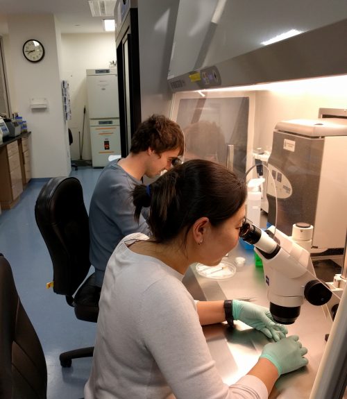
The first set of grafting took longer than I expected: even being very familiar with chick embryo manipulation, it was my first time trying to generate a chimera. It was late, I was exhausted and hungry crossing the narrow bridge back home, but I was also feeling an excitement and eagerness for the daybreak to see the results. As it turned out, our first grafted embryos looked more like a Picasso painting. I still think MoMa museum would love to exhibit our nightmarish sci-fi art. But a tweak here and there and we managed to keep the embryos alive and looking more… normal-ish! After that, it was a marathon. Besides the long hours in lab grafting, swayed by Brazilian forró songs and replenishing ATP with Iain’s hidden snacks, we also had to go through endless washing steps for in situ and long confocal imaging sessions.
And then, finally there it was! In the elongated structure emanating from the human cells we found expression of SOX2!! How awesome that could be?! To me, that was the mind-blowing moment, but of course I still had to hold my horses and wait for the in situ results of SOX3 probe to confirm our findings. SOX2 and SOX 3 were ectopically induced in chick cells that surrounded the human cells!! We had generated our very first chick-human chimera, our “Chuman”! Our results bring valuable insights into early human development.
This work was one of those “high risk, high reward” kinds of project. It could lead to an amazing discovery or could give us nothing. Gladly, with a wonderful teamwork, we got the reward!
1. Martyn, I., Kanno, T. Y., Ruzo, A., Siggia, E. D. & Brivanlou, A. H. Self-organization of a human organizer by combined Wnt and Nodal signalling. Nature 558, 132–135 (2018).


 (4 votes)