Microinjection of preimplantation mouse embryos
Posted by svanderweide, on 28 January 2013
 Hello, my name is Stephanie and I’m a graduate student in Dr. Amy Ralston’s lab at the University of California Santa Cruz. I just returned from a trip to Dr. Yojiro Yamanaka’s lab at McGill University in Montreal, Quebec. This trip was funded by the Development Travelling Fellowship from Company of Biologists. I highly recommend checking it out, receiving this grant was a great, hassle-free experience!
In my time at Dr. Yamanaka’s lab I learned how to synthesize and inject mRNA constructs into 2 and 8-cell mouse embryos. I also learned how to live image the embryos as they develop and analyze the data gathered from the imaging. During my visit I injected GFP and RFP mRNA for easy visualization of my injection success. I will be using these techniques back at UCSC to study the molecular regulation of the first lineage decision in the mouse embryo. The molecular mechanisms underlying the first asymmetries and subsequent lineage decisions in the mouse embryo are only beginning to be understood. I will be using microinjection to over-express a variety of intra-cellular signaling molecules and transcription factors and then assessing the fate of the injected cells.
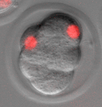
My time in Montreal was very cold! Coming from California I’m not adapted to living in subzero temperatures, and most days were below zero (Celsius). In fact, on the coldest day the high was -22 C, and felt even colder because of the windchill. It was great to visit French Canada though, very different from other regions in Canada. McGill University was very international and I met scientists from all over the world who are working there now. I’m glad to be back in California though, where the high today in Santa Cruz was 15C, well above zero. I’m also excited to get our injection system up and running and start collecting data. I included a picture of a 16 cell mouse embryo which I injected at the 8 cell stage. One cell was injected with H2B conjugated to RFP, marking the DNA, and that cell has subsequently divided. This was fun and very technically challenging to learn!


 (4 votes)