Please, show me your boundaries
Posted by Michalis Averof, on 18 November 2024
from Irene Karapidaki, Béryl Laplace-Builhé and Michalis Averof

What is this?
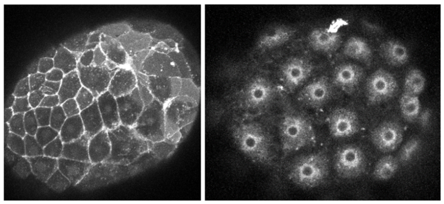
These are crustacean embryos injected with mRNA encoding a red fluorescent protein bound to membranes. On the left, the fluorescent protein is localised on the plasma membrane, on the right it is trapped in the endoplasmic reticulum and/or Golgi.
How was this image made?
These images were made while testing different membrane localisation tags in the marine crustacean Parhyale hawaiensis. One-cell stage embryos were injected with mRNAs encoding mScarlet3 fused to a Lyn tag, directing protein myristoylation and palmitoylation (on the left), or a signal peptide and the CD8 transmembrane domain (on the right). The embryos were allowed to grow for a day and then imaged live on a confocal microscope.
Why should people care about this?
Targeting fluorescent proteins to cell membranes allows us to visualise the shape and behaviours of cells in living embryos, as they build the body. See for example this amazing movie, showing the choreography of cells as they build sensory organs in the fish embryo: https://thenode.biologists.com/ready-steady-cooooooonga/research/
Can I do this in my favourite research organism?
The problem is that existing membrane-localising tags do not work equally well in all species. The SP-CD8 tag for example (on the right) gives good plasma membrane localisation in Drosophila, but gets stuck in the secretory pathway in our crustacean (Parhyale) embryos. In non-conventional model organisms, one needs to test several tags to find a good one.
To facilitate this process we generated a toolkit of 11 membrane-localising tags, which can be screened rapidly by microinjecting mRNA in your species of interest. Comparing results obtained in different species will help to identify tags that work well in a wide range of eukaryotes. If you are interested in trying the toolkit and joining our comparative screen, please get in touch.
Where can I find more about it?
Have a look at our preprint: https://www.biorxiv.org/content/10.1101/2024.11.12.623055v1
Check out other ‘Show and tell’ posts highlighting impressive images and videos in developmental and stem cell biology.


 (2 votes)