

Same cells, different gastruloids: what 2026 papers say about where embryo-model variability comes from
Posted by Shirley yang, on 20 November 2026
Small aggregates of mouse embryonic stem cells can break symmetry and organise an axis on their own, as first shown in Development in 2014. Given the right pulse of signals they lay out neural, mesodermal and endodermal derivatives along an elongating anterior–posterior axis, complete with collinear Hox expression. That is why gastruloids became so popular. Yet even under carefully controlled conditions, individual gastruloids differ substantially: some fail to develop, others lean towards one lineage.
Variation between structures is usually something to control for. Several papers from the past few months make it the object of study instead. Together they point to sources of variability at three stages (in the starting cells, during exit from pluripotency, and in the culture environment) and bring tools that measure one gastruloid at a time.
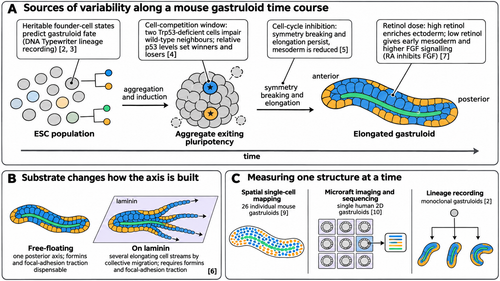
Figure 1. Where variability enters a gastruloid experiment, and how recent studies measure it. (A) Mouse gastruloid time course, from ESC population to elongated gastruloid, annotated with the sources of variability discussed in the text: heritable founder-cell states recorded with DNA Typewriter [2, 3]; a cell-competition window between primed pluripotency exit and early gastrulation, in which relative p53 levels set winners and losers [4]; cell-cycle inhibition, which leaves symmetry breaking and elongation intact but reduces mesoderm [5]; and retinol dose, which shifts the ectoderm/mesoderm balance through endogenous RA and FGF signalling [7]. (B) Substrate switches the morphogenetic mode: free-floating gastruloids form one posterior axis, whereas gastruloids on laminin form several elongating streams that require formins and focal-adhesion traction [6]. (C) Approaches that read out individual structures: spatial single-cell mapping of 26 individual gastruloids [9], single-gastruloid imaging and sequencing of human 2D gastruloids on microrafts [10], and lineage recording from monoclonal gastruloids [2]. Bracketed numbers match the labels in the figure and link to the papers. Schematic, not to scale. Schematic created with Scientific Figure.
Heterogeneity starts before induction
The most direct test comes from Regalado and colleagues in Cell, who made monoclonal gastruloids, each grown from a single mouse ESC. Some failed to develop, some were biased towards mesodermal or neural fates, and some contained cell types atypical of conventional polyclonal gastruloids. Using DNA Typewriter, a molecular recorder built on sequential genome editing, the authors found heritable fate biases that arose before gastruloid induction: more closely related founder cells gave more similar gastruloids. The fate-predictive differences sat in gene expression and chromatin accessibility, including at imprinted, methylation-sensitive loci linked to developmental potency. The logic of a fluctuation test is simple: if outcomes cluster by lineage, the variation was already present in the starting cells.
A related bioRxiv preprint from the Zernicka-Goetz lab, working with the same lineage recorder, followed the same idea into the embryo. Combining DNA Typewriter with tetraploid complementation, the authors report that loss of developmental potential in mouse embryos is progressive and asynchronous, with several lineages showing detectable fate bias before morphological diversification at gastrulation. In a stem-cell-derived embryo model, cells with the same transcriptional identity could arise from different lineage relationships. It is a preprint and not yet peer reviewed, but it lines up with the monoclonal gastruloid result: cells that look alike may not share a history.
A narrow window for cell competition
If some founder cells are “better” than others, does the aggregate sort that out? Frenster and colleagues, in Nature Cell Biology, built mosaic mouse gastruloids to ask how cell competition plays out in 3D. Just two Trp53-deficient supercompetitor cells were enough to impair the growth of their wild-type neighbours through mitochondrial apoptosis. Competition was restricted to the transition between primed pluripotency and early gastrulation. Using heterochronic gastruloids and p53 degrons, they showed that both winners and losers have to be in that permissive stage, and that relative p53 protein levels decide the outcome. So the early aggregate is not a passive mixture, and when cells become unequal matters.
Proliferation is a second variable. Leonardi, Paychère and Naef (iScience) inhibited the cell cycle in mouse gastruloids: despite near-complete growth arrest, they still broke symmetry, elongated and specified germ layers. What changed was the balance, with delayed differentiation and fewer mesodermal cells. Proliferation differences between cell types only partly explained this; the authors conclude that cell-cycle dynamics also tune lineage-specific differentiation timing and efficiency. Morphology can look fine while composition drifts.
Shape and composition follow the culture conditions
Two studies show how much the environment steers the outcome. Serrano Nájera, Delahaye and Steventon, in PNAS, plated mouse gastruloids on laminin and compared them with free-floating ones. Free-floating gastruloids built a single posterior axis. On laminin they flattened, broke symmetry several times and produced several independently elongating cell streams with anterior–posterior polarity, through collective cell migration. Formins and focal-adhesion traction were needed for elongation on laminin but were dispensable in suspension. Formin inhibition blocked elongation on laminin without detectable transcriptional changes. Laminin also biased cells towards posterior migratory fates, while Hox expression stayed similar.
Medium composition matters in the same way. Yildiz and colleagues, in Development, replaced added retinoic acid with its precursor retinol in a retinoid-free medium, so that cells made RA themselves. High retinol enriched ectoderm, while low retinol led to early mesoderm specification and higher FGF signalling. They also confirmed that RA signalling inhibits FGF in this system.
Human gastruloids, reported in Nature in 2020 as a model of early anteroposterior organisation, bring their own protocol questions. Azami, Patton and Nichols, also in Development, went through the protocol one parameter at a time across four hPSC lines from the UK Stem Cell Bank. They switched to home-made N2B27, cut activin A to 15% of the original concentration, titrated initial cell density, delayed the brief GSK3-inhibitor exposure until aggregation began, and added a TGFβ inhibitor, which further improved how often the three germ layers regionalised as expected. The stated aims are efficiency, cost and reproducibility, and the paper is a reminder of how many small decisions sit inside the word “protocol”.
Measuring one gastruloid at a time
Much of this is invisible in pooled samples. Triandafillou and colleagues, in eLife, produced spatially resolved single-cell transcriptomes for 26 individual mouse gastruloids. Cell-type composition and tissue-scale organisation were largely consistent between them, but meso-scale patterning of specific cell types varied. Posterior cell types formed distinct, organised clusters; anterior cell types were more disorganised. They also confirmed that neuromesodermal precursor differentiation proceeds as a continuous, spatially coordinated process.
For human 2D gastruloids, Jan and colleagues (APL Bioengineering) built a microraft-based assay to screen, sort and sequence individual living gastruloids carrying germ-layer reporters. Time-series imaging analysed with self-supervised deep learning identified distinct patterning trajectories, and single-gastruloid transcriptomes clustered by batch, with subclusters within batches. The authors are candid that their sample was too small to link imaging phenotype to expression, and frame the study as proof of concept.
What follows for people using these models
Taken together, these papers suggest a few practical points. Some of the variation between gastruloids is heritable from the starting cells. The early aggregate goes through a window in which relative cell fitness shapes who contributes. Composition can shift while gross morphology looks normal, whether the cause is a slowed cell cycle or the retinol dose. Substrate and medium can switch the morphogenetic mode altogether. Batch structure shows up even in single-gastruloid data, which is why Jan and colleagues call for careful evaluation of reproducibility in human in vitro systems. Reporting per-structure measurements, and how many structures were excluded and why, seems a reasonable response. And the mosaic and monoclonal designs above show the other side of the coin: once variability can be traced to a cause, it becomes another way to ask how development stays robust.
AI tools were used to help search the literature and draft this post. All claims were checked against the linked papers.
Get involved
Create an account or log in to post your story on the Node.
Sign up for emails
Subscribe to our mailing lists.