See for yourCelLf
Posted by Joachim Goedhart, on 18 August 2017
See for yourCelLf
“Forget the textbook picture” is what I proclaim when I teach master students in a course on Cell biology and Advanced Microscopy. Although the textbook is a fantastic resource for teaching, it largely fails to convey the complexity of cells, including their size, dynamics and structure. To fully appreciate the intricacies of cells, one needs to get in touch with the material. This involves going to the lab, prepare samples, observe the cells through a microscope and acquire images.
One of the most fun experiments that I supervise during a cell biology course deals with the size and shape of organelles. In this experiment, the organelles of cells are labeled with fluorescent markers that are genetically encoded on a plasmid. The students have to examine five different samples and infer which organelles are highlighted in each of those samples. The eagerness of getting it right motivates the students to carefully observe the cells and compare between different patterns of localization. Once every group of students has reported their findings on a white board, the results are discussed. The public reporting and discussion brings an element of competition to this experiment and it adds to the motivation to correctly identify the organelles.
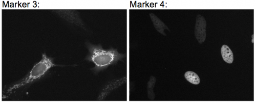
To perform this experiment, we use plasmids that encode different ‘cellular markers’, each capable of highlighting an organelle in a mammalian cell*. The plasmids are isolated from bacteria by the students in a blind fashion, i.e. the students do not know what organelle is labeled by the markers they isolate. The students transfect the cells with the unknown markers (we use HeLa cells for ease of culture and robust transfections). The next day, the students are given the task to prepare samples, observe cells with the different markers and identify them**. Many iterations of this experiment are imaginable, depending on the time and equipment*** that is available. For instance, endogenously tagged human stem cells (e.g. from the Allen cell collection) can be used to make the assignment more relevant from a biomedical perspective.
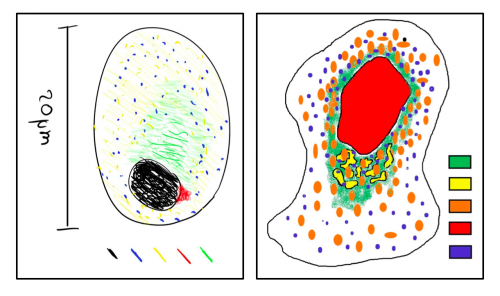
In addition to identifying the organelles that are labeled, I ask the students to compare their results to a textbook image and generate an improved picture based on their observations. Altogether, this assignment is fun and I’m convinced that observing fluorescently labeled organelles (or other structures) in cells results in a better understanding of cellular function. I’d love to hear about your experiences in teaching cell biology and the experiments that students perform to learn about the intricacies of cells.
—————
*We have generated and reported several markers that are suitable for this purpose. These and others are available from addgene.
**I decided not to disclose which markers we use for obvious reasons. If you would like to know which markers we use, or if you would like more background on the experiment feel free to contact me by email or a tweet.
***The possibilities to implement a practical course in cell biology will depend primarily on the equipment that is available. At our university, we are lucky enough to have 14 basic fluorescence microscopes available for teaching 40 students at a time (160 in total). These microscopes are equipped with basic fluorescence filter sets (DAPI/FITC/TRITC) and simple CCD cameras.
 (6 votes)
One thought on “See for yourCelLf”
Leave a Reply
Get involved
Create an account or log in to post your story on the Node.
Sign up for emails
Subscribe to our mailing lists.
Read the latest Development issue
Most-read posts in May
- From comparison to mechanism: decoding heart regeneration
- A day in the life of a Reviews Editor (at Development)
- What does a Reviews Editor do?
- New evo-devo textbook ‘Eco-Evo-Devo: The Environmental Regulation of Development, Evolution, and Health’
- preLighters’ choice – A curated selection of recent preprints
This is a wonderful lab experiment for students and I am certain I would have loved it!
We did a similar one in genetics, having to figure out a genetic cascade from embryo mutants from Janni Nusslein-Volhard/Eric Wieschaus collection!
Thank you!