Some of the things we have done during these two weeks
Posted by Lucia Prieto Godino, on 27 October 2011
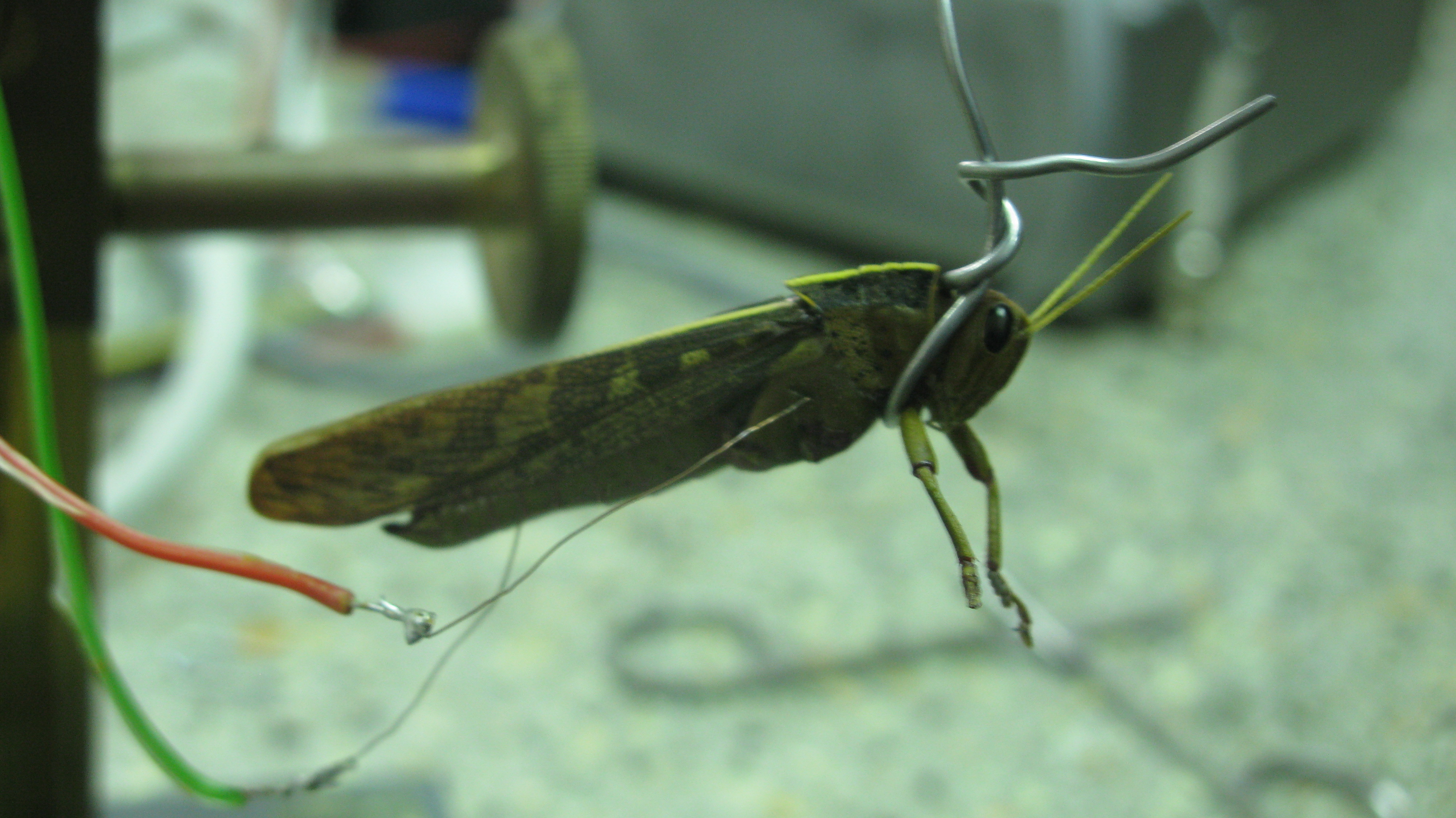
We have now finished the first two weeks of the course. Over these two weeks the students have learned about Drosophila as a model organism and how to set-up a Drosophila lab. They have learn about the genetics of Drosophila, which took quite some time, but now I am sure it is almost like their mother tongue. They have also learned basics concepts in neuroscience, such as the nature of neural impulse, and basic concepts on how neurons work to produce adaptive behaviors. During the practicals among other things they have learned how to do muscle recordings of neuronal activity with inexpensive amplifiers. They have recorded from the legs and wing muscles of grasshoppers (see picture 1)! and observed under the microscope a multitude of different insects, which all together helped them to appreciate in the practice the nature of the neural impulse and the diversity of sensory and motor systems used by insects.

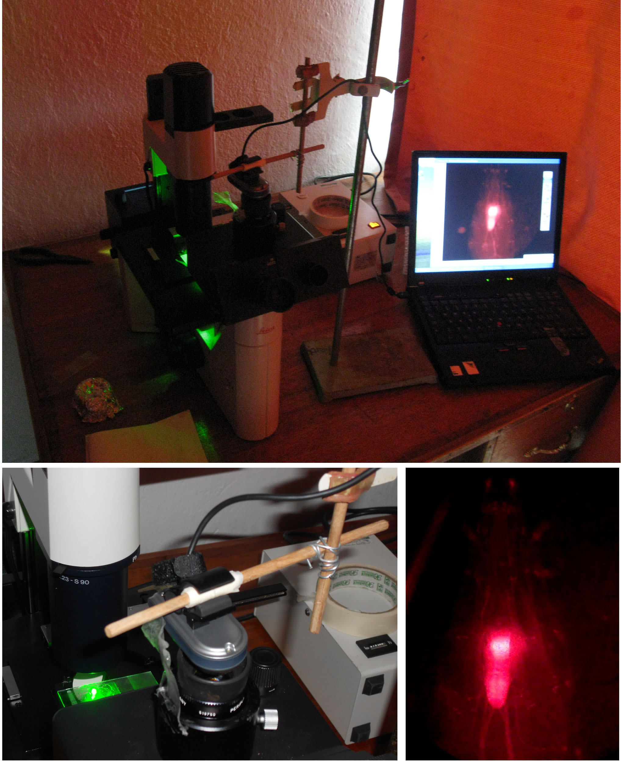
The students also now know how to collect virgins for their fly crosses, and how to dissect brains out of Drosophila larvae, and look at them under the fluorescent microscope. We have managed to install a webcam on the fluorescent microscope, so that the students can take pictures of the fluorescent preparations that they look under the microscope. The images resulting from this system are actually much better than we expected (see picture 2).

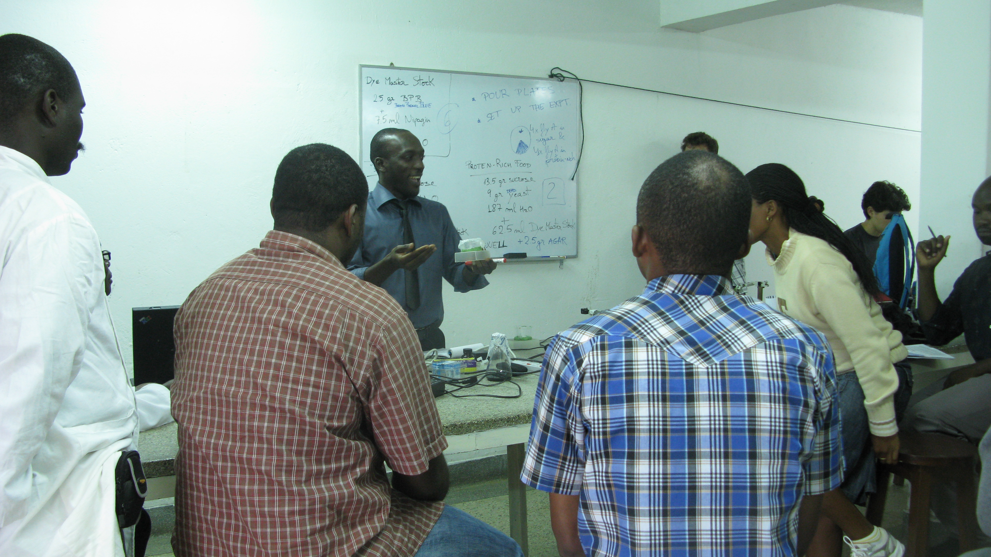
The students have learned about mechanosensory, chemosensory, visual and motor systems during the theoretical lectures, and they have looked at the wild type behaviours of flies and other big insects during the practical sessions (see picture 3).

They have also performed inexpensive cutting-edge neurogenetic experiments on genetically modified larvae. It is being intense but the effort is worthwhile, now they are ready for the lab work of the last week, during which they will need to apply everything they have learned!


 (3 votes)