Synchronicity: Laser-targeted ablation timed with zebrafish embryonic heart cycle
Posted by Cindy Rodzen, on 29 July 2013
![]() Matrone G, Taylor JM, Wilson KS, Baily J, Love GD, Girkin JM, Mullins JJ, Tucker CS, Denvir MA. Laser-targeted ablation of the zebrafish embryonic ventricle: A novel model of cardiac injury and repair. Int J Cardiol. 2013 Jul 17. doi:pii: S0167-5273(13)01117-0. 10.1016/j.ijcard.2013.06.063. [Epub ahead of print] PubMed PMID: 23871347 (Open Access Article)
Matrone G, Taylor JM, Wilson KS, Baily J, Love GD, Girkin JM, Mullins JJ, Tucker CS, Denvir MA. Laser-targeted ablation of the zebrafish embryonic ventricle: A novel model of cardiac injury and repair. Int J Cardiol. 2013 Jul 17. doi:pii: S0167-5273(13)01117-0. 10.1016/j.ijcard.2013.06.063. [Epub ahead of print] PubMed PMID: 23871347 (Open Access Article)
In a study published July 17, 2013 in the International Journal of Cardiology on line, researchers found that “laser-targeted injury of the zebrafish embryonic heart is a novel and reproducible model of cardiac injury and repair suitable for pharmacological and molecular studies.”
The scientific team, led by Dr. Martin Denvir, College of Medicine and Veterinary Medicine, University of Edinburgh, UK, undertook this study to learn more about how the embryonic zebrafish heart responds to injury as compared to the adult zebrafish heart, which demonstrates a remarkable capacity for regeneration.
At the “heart” of this study was the XYClone infrared laser with RED-i target, from Hamilton Thorne Inc.. The researchers produced targeted and highly localized injury to the embryonic heart by synchronizing the XYClone laser pulse with the cardiac cycle. By using custom software, they were able to apply the laser pulse only at a specific user-designated phase of the cardiac cycle, which allowed targeting of just the embryonic heart ventricle.
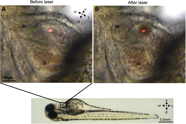
Zebrafish embryos 72 hpf (lower panel)were used for all experiments of laser injury. The laser pulse was delivered to the area of the ventricle indicated by the red dot (Panel A) and resulted in a clear burn-mark at the point of injury (Panel B), see also supplementary movie 1 (V – ventricle, BA – bulbus arteriosus, At – atrium). Position of the embryo is marked by compass lines (c-caudal, cr-cranial, d-dorsal, v-ventral)
Cardiac arrest and cessation of tail blood flow demonstrated the immediate injurious effects of the laser. In addition, cell death and apoptosis resulted in loss of cardiomyocytes. A significant decrease in heart function was observed, yet, by 24 hours post-lasering, complete recovery occurred. The study results showed, for the first time, that a proliferation of new cardiomyocytes drove the functional recovery of the lasered embryo heart ventricle. It also appeared that the laser injury itself stimulated the proliferative process.
In the discussion, the authors note many advantages to using the laser model, including the rate at which the individual zebrafish embryos may be processed, the reproducibility, the ease of testing pharmacological and genetic interventions, and the ability to create regional damage similar to that which occurs from ligation of the coronary artery in mammals.
Supplemental videos:
Movie 1: Laser pulse injury (without synchronisation) of the zebrafish embryonic heart ventricle at 72 h post-fertilization– A single laser pulse, using the XYClone Laser Ablator, to the ventricle of a zebrafish embryo (72 hpf) results in instantaneous cardiac injury associated with marked bradycardia and gradual recovery of cardiac rhythm over the next few minutes. A laser burn-mark is clearly seen in the wall of the ventricle. This is an example where there is a clear view of non-overlapped cardiac chambers.
MOVIE 2: Laser pulse injury using the synchronization software of the zebrafish embryonic heart ventricle at 72 h post-fertilization. In this example, atrium and ventricle are overlapped. Attempting to injure the ventricle with a non-synchronized laser system would result in damage to adjacent structures. Synchronizing the laser pulse with the cardiac cycle allows highly precise and targeted injury to the ventricle at end-diastole and consequently minimizes damage to surrounding structures.
Note: The author, Cindy Rodzen, is affiliated with Hamilton Thorne Inc.
 (1 votes)
Get involved
Create an account or log in to post your story on the Node.
Sign up for emails
Subscribe to our mailing lists.