The 2012 UK National Xenopus Conference
Posted by Victoria Hatch, on 10 April 2012
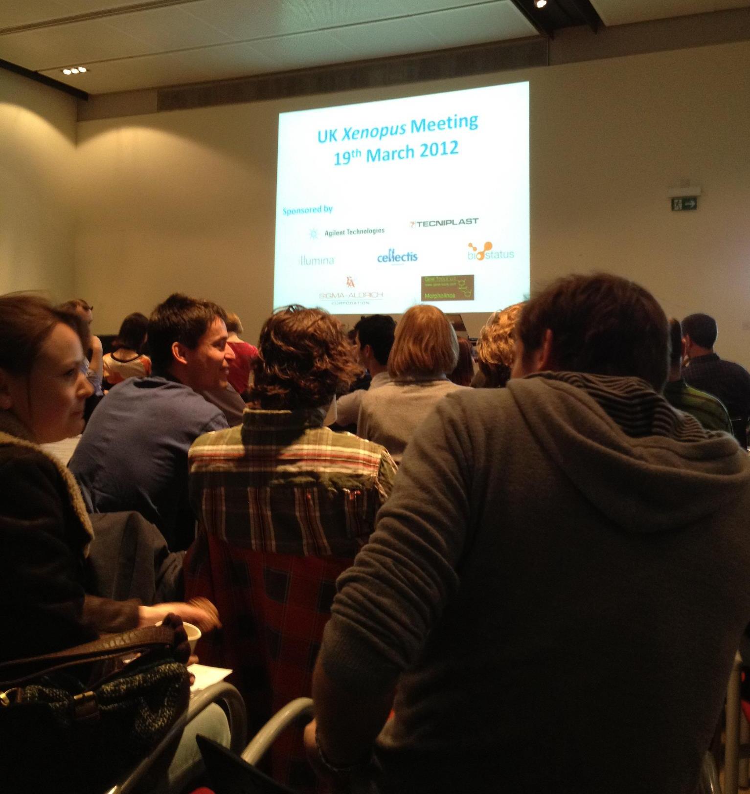
The UK national Xenopus conference is an annual event held to discuss the exciting and extremely varied work carried out across the UK using the African clawed frog (Xenopus) as the model organism of choice. The event provides opportunity for PhD students and Postdocs from Xenopus labs round the UK to present their data to experts within the Xenopus community, a community which can easily be described as passionate and welcoming to all new members. This year’s meeting was held on the 19th of March at the Wellcome Trust Sanger Institute (Cambridge, UK). This institute, opened in 1992, is famous for its substantial participation in the sequencing of the human genome. As a single institute it alone contributed the most sequencing data for what is now considered the gold standard human genome sequence. The meeting was held in a conference room lined with stands for some of the many sponsors of the event. These included companies such as sequencing giants Illumina, morpholino pioneers Gene Tools and many others including Techniplast, Sigma-Aldrich, Agilent Technologies, Biostatus and Cellectis Bioresearch.

The talks kicked off with a welcome from Derek Stemple, a senior investigator at the Sanger Institute who is currently using Zebrafish and Xenopus tropicalis as human disease models as well as being involved in the Zebrafish genome sequencing project. His welcome outlined the history of the Sanger Institute and gave a strong message of the importance it has played in revolutionising genome sequencing over the last 20 years and its continuing research into genomics and human disease models. This posed as a great introduction for the first talk by Amanda Hall who is currently working on the Xenopus tropicalis mutation resource project at the Sanger Institute. This project aims to create Xenopus knockouts which can be used to study various disease models. Invitro sperm ENU mutagenesis has been used to generate 6000 F2 mutant individuals the progeny of which can be maintained as genomic DNA libraries and frozen sperm for verification once positive phenotypic analysis is carried out on an F3 carrier population. The genomic library will undergo a reverse genetic screen known as TILLING to indentify 175 preselected mutations. This project can greatly benefit the Xenopus community as all mutations will be made available to view on an online database on the Sanger Institute website. Also all mutant sperm and living animals can be made available for use in further research.
The second talk of the day was by Anita Abu-Daya who is currently working in Lyle Zimmerman’s lab at the National Institute of Medical Research (NIMR) in London. Lyle’s lab has collected a variety of weird and wonderful Xenopus tropicalis mutations over the last few years by the means of gynogenetic screens. In her talk, Anita spoke of a mutant called Whitehart, which after phenotypic analysis was identified to have no circulating blood. Genetic analysis of Whitehart revealed it to have a mutation resulting in a premature stop codon in the Smad 4.1 gene, the mediator Smad of both TGFβ and BMP signalling. But if the embryos lack such an important Smad then why is the phenotype not more extreme? This was deduced to be down to redundancy by the similarly expressed Smad 4.2. This work in summation gave a potential role for BMP signalling for the maintenance of blood. The third talk was given by Nick Owens from Mike Gilchrists lab also at the NIMR. His talk outlined the importance of biological variability when using techniques such as RNAseq. To prove this importance he uses two inbred mothers whose eggs are fertilised with the same father. 8 pools of 20 embryos were collected from both parent combinations to undergo sequencing. His data generally shows the more replicates the better to avoid biological variability. Three replicates will always give more reliable results than two.
The next set of talks included one from Tom Bates of Esther Bell’s lab (Kings College London). His work showed a novel TGFβ inhibitor, Coco, to be expressed in the animal pole of gastrula stage embryos. Knock down of Coco by host transfer techniques and morpholino injection showed a loss of dorsal tissues. His work could be summarised by a model suggesting Coco to be regulating germ layer specification via its inhibition of TGFβ in the animal pole. Siwei Zhang of Enrique Amaya’s lab (University of Manchester) spoke about the role of Fezf2/TLE4 in diencephalon development. He showed Fefz2 to be expressed during Xenopus neurulation and in the forebrain of tailbud and tadpole stage embryos. mRNA injection of Fefz2 resulted in dorsal anteriorized embryos leading to a hypothesis of Fefz2 regulating Wnt signalling in the diencephalon area. Next up was Natalie Gibb from Stefan Hoppler’s lab (University of Aberdeen). She demonstrated a role for Sfrp1 in promoting myocardial differentiation. Her talk contained some stunning images and videos demonstrating the ability of Sfrp1 to modulate the size of the developing heart. Coinjection of Sfrp1 and Wnt6 rescued the dual axis phenotype usually associated with Wnt overexpression. This indicates a role for Sfrp1 in modulating Wnt signalling during myocardial development. The last talk of this set was from Neil Roberts (University of Manchester). His work used Xenopus as a disease model to characterise mutations which may result in the unusual disease, Urofacial syndrome a human disease with symptoms including bladder dysfunction and abnormal facial expressions. His work, funded by Kidney Research UK, indicates potential for mutations in the Heparanase-2 gene to be causative of the disease.
Between talks a delicious buffet lunch was supplied which allowed all conference attendees to sit outside and enjoy the outrageously beautiful day upon which the conference took place. With the sun shining everyone moved outside to continue their discussions and meet new people within the community. Also at this time a tour was provided of the Sanger Institutes very own Illumina HiSeq equipment allowing certain attendees to see these powerful machines in action. This also gave the opportunity for us to talk to some of the sponsors. One which I found particularly interesting was Biostatus whom have developed what they are calling CyGEL, a thermoreversible gel which can be used to immobilise living organisims such as the Xenopus allowing live cell imaging.

The next round of talks started with Jerome Jullien of John Gurdon’s lab. He demonstrated genome wide reprogramming of somatic nuclei by the Xenopus oocyte transcription machinery. Nuclear transfer was shown to increase Serine 2 phosphorylation found on RNA polymerase II, a phosphorylation state associated with transcription elongation. Clara Collart of Jim Smith’s lab (NIMR) gave the next talk on the role of YRNAs during midblastula transition (MBT). YRNAs are expressed maternally and their expression increases after MBT. Mopholino knockdown of specific YRNAs results in normal looking embryos until MBT which is known to be the onset of transcription. All previous transcription occurring in the embryo is maternal. Clara demonstrates that YRNAs are capable of interacting with chromatin after MBT to promote the onset of DNA replication in Xenopus. Hugh Woodland (University of Warwick) finished this set of talks with a rather puzzling and insightful presentation demonstrating the relationship between oocyte and follicle cell RNA localisation. RNAs are known to exchange via transport pathways between the oocyte and macrovilli/nanotubules extending into the surrounding follicle cells. Conversely, Hugh’s data provides evidence for particles in the oocyte nanotubules to represent an RNA transport pathway from follicle cells to the oocyte. This suggests that follicle cells may contribute to stored egg RNAs. To solve this conundrum he has transplanted primordial follicle mesoderm from Xenopus borealis into Xenopus laevis to see which species RNA will be present in the follicle cells and oocytes. The problem is he still has a long time to wait for these frogs to fully mature.
The last set of the day started off with Eric Theveneau from Roberto Mayor’s lab (UCL). He presented the importance of placodes, found in the gaps between branchial arches, in modulating neural crest migration. He uses Sdf1 as a marker for the placodes to demonstrate an intrinsic attraction of neural crest cells towards Sdf1 positive cells leading to efficient co-ordination of these cell types during cranial morphogenesis. Roberto Paredes (University of Manchester) next spoke of his research into the behaviour of myeloid cells after injury. He has developed an effective method to visualise myeloid cells in vivo demonstrating a fast response by neutrophils in response to injury and a comparatively slower response by macrophages. The last talk of the day was given by Matt Guille (University of Portsmouth) who when not conducting his own research is an indispensible member of the Xenopus community for his work in managing the European Xenopus Resource Centre in Portsmouth. Today however, he was not talking about the centre but his own research on histone function in early Xenopus development. The specific histones H2A.Z1 and H2A.Z2 were the primary focus of the talk. These are implicated to be involved in transcription activation and differ by only 3 amino acids. They give similar expression patterns found at several stages of Xenopus development. Knock down of H2A.Z1 shows a loss of Lmo2, Tbx, Hex and Brachyury shown by insitu hybridisation and animal cap experiments.
One of the most important parts of the UK national Xenopus conference is the community discussion at the end. This gives anyone the chance to voice concerns over all manner of topics including the Portsmouth resource centre, funding and even problems arising with scientific techniques. The key points made in this discussion included the announcement of the opening of the American Xenopus resource centre which can be found at Woods Hole, Massachusetts. A general update of the resources available at the Portsmouth centre. Finally, a suggestion to all Xenopus researchers to keep a careful log of antibodies they have previously used and found to be successful. This kind of information could save Xenopus researches a lot of valuable time and money by producing an accessible database of these antibodies. The day ended with the usual drinks and nibbles and for me a short drive home to Norwich. I had a fantastic day at the conference and would strongly recommend attending it next year if you are working in the Xenopus field. I personally will be eagerly anticipating what new and exciting research next year’s meeting will bring.


 (10 votes)