This month on the Node – January 2013
Posted by the Node, on 31 January 2013
Let’s start this monthly summary with two fantastic posts that were left out of last month’s roundup due to the holiday period (and scheduled posts).
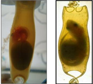 Idoia Quintana studies shark brain development in Spain, and travelled to Scotland recently to learn how to work with mouse brains, so she could do some cross-species comparison.
“At the beginning was tricky, working with mice was a big challenge for me; but at the same time was amazing to do research in a species in which several optimized techniques are available. Particularly I enjoyed learning slice culture techniques and I hope to have time to implement them in shark embryos and perform some axon guidance experiments upon my return.”
Benjamin Coyac attended the UPMC/Curie Institute International Course in Developmental Biology, and described what it was like to study in Paris for several weeks with students from around the world.
“The intensity of the program enabled us to get to know each other very quickly. Little talks about science or our lives as international students began to build a common experience in our shared interest in developmental biology. By the end of the program, not only we had acquired scientific skills in theory, methods and practicals, but also we became friends and started to build a strong network of future developmental scientists.”
And now on to the January posts.
Mouse development
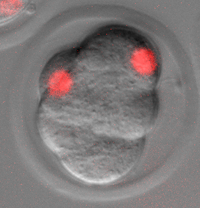 Stephanie Vanderweide travelled from California to Montreal in the middle of the Canadian winter to learn how to microinject 2-cell and 8-cell mouse embryos in Yojiro Yamanaka’s lab, as part of a project to study the molecular mechanisms involved in the first lineage decision of the developing mouse embryo.
Heather writes about a technique developed in the Niswander lab, where she’s a student. The lab set up a confocal-based live imaging system to visualize mouse embryo development in real time.
“We have been using this system to study neural tube closure, but there are many other tissues and organs that develop during these time periods (E8.5-E10.5) that are amenable to imaging including the heart, face, limbs and neural crest. By using tissue-specific Cre- recombinase reporter strains, the behavior of individual cell types can now be observed in real time in the early mammalian embryo.”
Woods Hole Embryology course
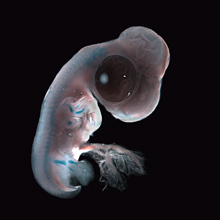
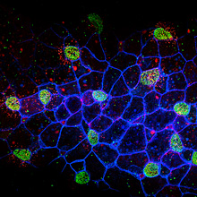
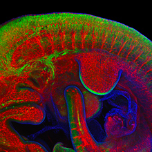
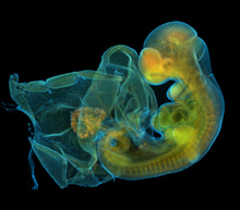
The application period for the 2013 Woods Hole Embryology Course has been extended to February 8. If you’re accepted to the course, we may see your images on the Node in the future: this is the course that produces the gorgeous images that Node readers have selected for Development covers. Speaking of which, the first round of images from the 2012 course are now up!
Publishing discussions
Finally, we’ve covered two very different discussions related to scientific publishing: peer review and the secrets behind methods sections.
-Katherine Brown wrote a post responding to an earlier discussion, about the future of peer review – both in general and in the specific case of Development.
-Eva followed the #overlyhonestmethods hashtag on Twitter, where within a few days hundreds of scientists shared the true stories behind their methods sections.
Also on the Node
-Lots of new job ads
–Hope for Huntington’s
–Top posts of 2012


 (1 votes)