Transparent mouse embryos and hematopoietic cell clusters
Posted by Erin M Campbell, on 8 November 2010
I was lucky in graduate school and my postdoctoral research—I was a microscopist working on a transparent organism (C. elegans). Some microscopists don’t have that luxury, but have developed amazing techniques in order to visualize development in organisms such as mice. In the November 1 issue of Development, Yokomizo and Dzierzak use a technique that makes an entire mouse embryo transparent and ready for high-resolution confocal microscopy, and they describe a comprehensive analysis of hematopoietic cell clusters.
Hematopoietic cell clusters are bunches of cells found on large blood vessels in mouse embryos and play an important role in the development of the adult blood system. Based on earlier reports, it was believed that hematopoietic cell clusters contained hematopoietic stem cells (HSCs), which give rise to many blood cell types. By using a whole-mount transparency method and 3D reconstructions of a mouse embryo, Yokomizo and Dzierzak constructed a temporal and spatial map of all hematopoietic clusters. The number of clusters peaks at embryonic day 10.5, and the clusters are found in specific subregions on vessels. In addition, Yokomizo and Dzierzak show that some clusters do contain HSCs and progenitor cells, confirming the pivotal role of cell clusters in the formation of the adult blood system.
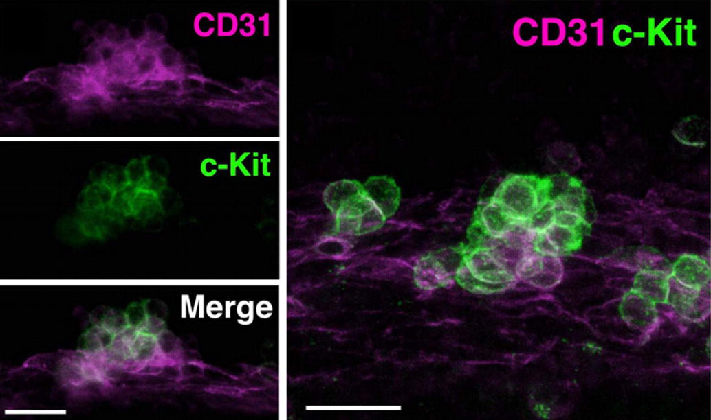
Images above show hematopoietic cell clusters on the aorta in mouse embryos. CD31 (magenta) is expressed by both the endothelial cells on the aorta and clusters cells, while c-Kit (green) is expressed only by cluster cells. The high magnification view of the cluster on the right shows how closely the clusters are situated next to the endothelium of the aorta.
Yokomizo, T., & Dzierzak, E. (2010). Three-dimensional cartography of hematopoietic clusters in the vasculature of whole mouse embryos Development, 137 (21), 3651-3661 DOI: 10.1242/dev.051094


 (5 votes)