When did our flexible and lubricated joints evolve?
Posted by Neelima Sharma, on 30 April 2025
[Behind the paper story of “Synovial joints were present in the common ancestor of jawed fish but lacking in jawless fish”.]
Synovial joints are marvels of biological evolution where two bony segments with curved articular geometries move relative to each other to produce motion and function [1]. In contrast, mechanical joints such as those present in door hinges, machines, and locomotives, also articulate but with simpler geometries, more limited directions of motion, a constant center of rotation, and no natural lubrication. How synovial joints develop fascinated me, especially the role of muscle contraction in joint morphogenesis. Multiple studies over several decades have shown that if the muscles of tetrapod embryos such as mice and chicks are paralyzed, many of their synovial joints remain fused and do not cavitate [2, 3]. I wanted to study the role of embryonic muscle contraction in shaping the articular surface geometries, as learning this relationship may harbor clues on the structure-function relationship of these joints and help design better prosthetics.
To understand the process of joint morphogenesis, I started my postdoctoral research in Neil Shubin’s lab and shifted gears from working in applied mathematics and biomechanics during my PhD to evolutionary developmental biology. Using dissection studies, I identified skates and sharks as the potential model organisms for studying synovial joint development, as they exhibited a large range of motion at the pelvic and jaw joints, and were comprised of striking articular geometries. However, I quickly learned that we did not know whether elasmobranchs have synovial joints at all. Upon deeper investigations, I learned that the situation was even more obscure; we did not know when synovial joints originated in the vertebrate phylogenetic tree! Where did our movable joints come from? I got intrigued by this question and chose to investigate the early evolution of synovial joints before delving into the processes of articular geometry morphogenesis.
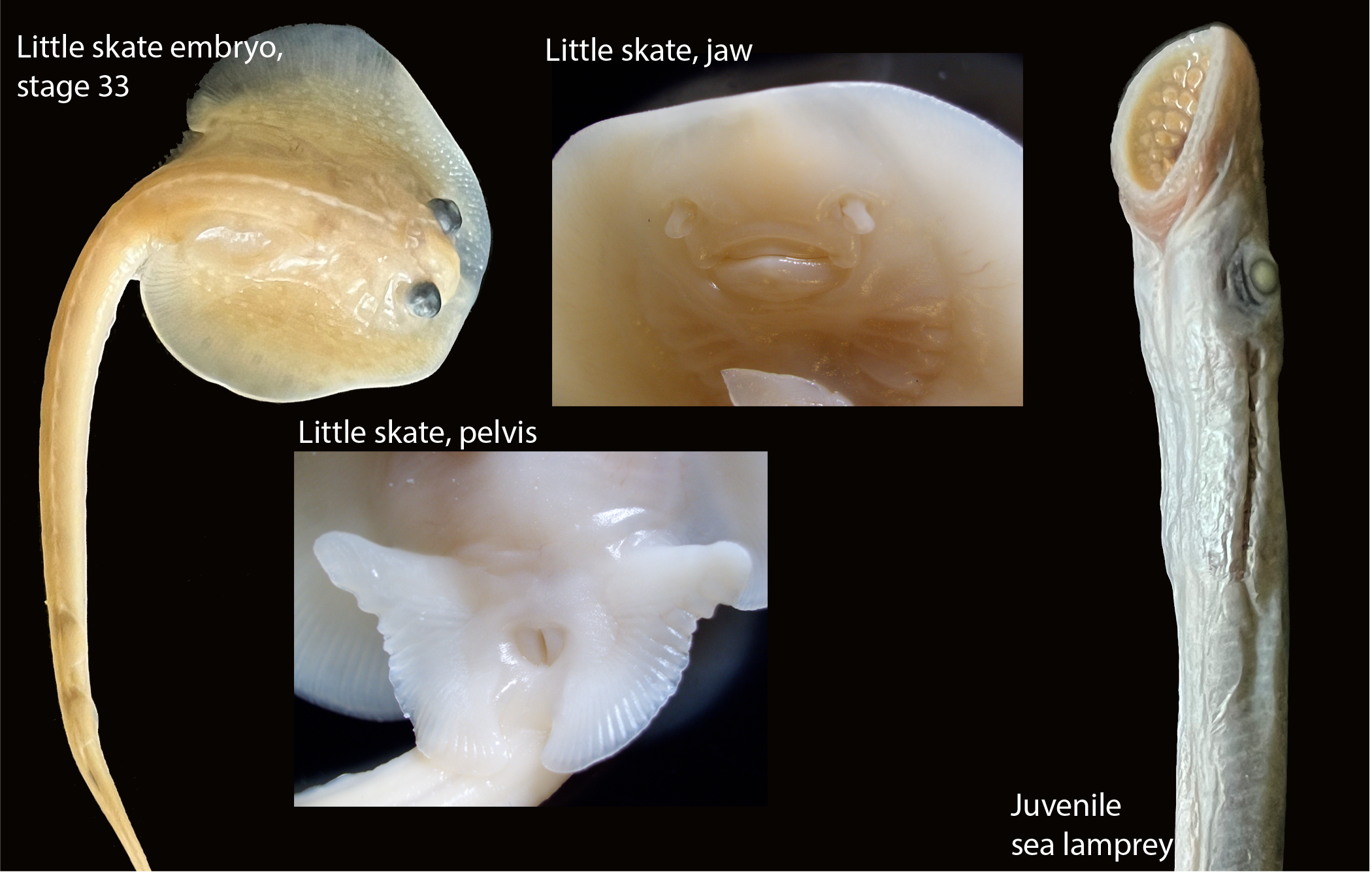
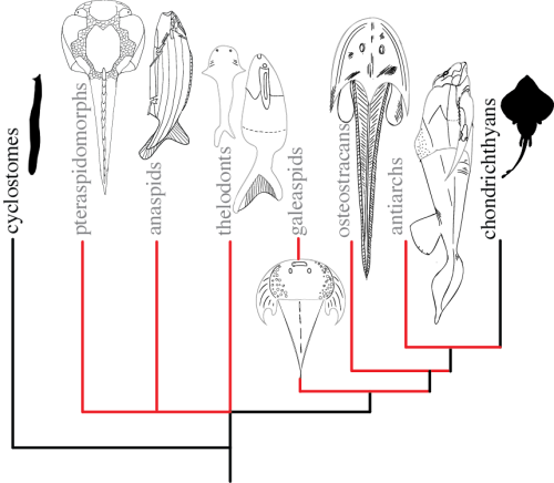
Apart from a couple of histological studies from the 1950s in elasmobranchs and chimeras that suggested the presence of cavitated joints, no deeper molecular and developmental investigations had conclusively shown the existence of synovial joints in these groups [4, 5]. To find the origin of synovial joints, I rolled up my sleeves and delved into learning the tools, techniques, scientific thinking, and methods of molecular and evolutionary developmental biology. A recent study out of Gage Crump’s laboratory at the University of Southern California showed how teleosts like zebrafish have synovial-like morphology in their jaw and pectoral fin joints [6]. I identified the two vertebrate clades that phylogenetically precede teleosts with extant representatives, cyclostomes and elasmobranchs, for whom the joint morphology was not clearly understood (Figure 1). I collected adult and juvenile specimens of lamprey and hagfish belonging to the jawless cyclostomes, and little skates and bamboo sharks belonging to jawed elasmobranchs, and performed micro-CT scanning and histological studies to show that little skates and bamboo sharks had cavitated joints. However, we did not find any evidence of cavitated joints in cyclostomes like lamprey and hagfish.
In the absence of cavitated joints, it is infeasible for synovial joints to exist because they need articulating surfaces and function by relative sliding. Therefore, we concluded that cyclostomes do not have synovial joints and focused solely on testing whether the cavitated joints of little skates, belonging to chondrichthyans, a constituent group within elasmobranchs, are synovial-like. Chondrichthyans have a cartilaginous skeleton, and therefore, the conventional definition of synovial joints, described as bony elements covered by a layer of articular cartilage, does not apply. However, if little skates have synovial joints, we expect their articular cartilage and subarticular regions to be morphologically and developmentally similar to tetrapods. To test this, I investigated the collagen proteins that composed the articular and subarticular cartilage in the little skate jaw and pelvis joints. Similar to tetrapods, the subarticular cartilage in the little skates was rich in collagen-II, and the developing articular cartilage in collagen-I (Figure 2).
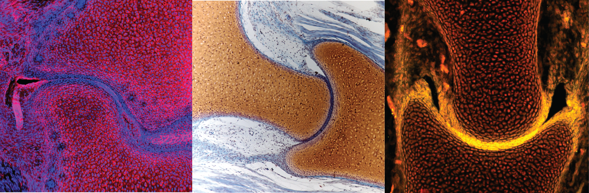
To test whether the joint cavity in little skates is lubricated, I tested whether any lubricating proteins are present in their articular cartilage (Figure 2). I performed in situ hybridization to locate the expression of lubricin, a key lubricating protein secreted by the articular cartilage and present in synovial fluid, as also shown in the articular cartilage of zebrafish [6]. As a newcomer lacking experience in molecular biology techniques, I did not understand how difficult in situ hybridization experiments can be. After multiple failed experiments for locating the expression of lubricin in little skates, I found a new respect for the experimental endeavors and gave up on looking for lubricin (if somebody is successful, please let me know!). Instead, using immunostaining, I showed the presence of other proteins such as aggrecan and hyaluronic acid receptors in the articular cartilage, also a part of the lubrication assembly [7, 8]. Furthermore, I showed that similar developmental pathways relying on Wnt and BMP signaling underlie joint development in little skate and tetrapods. Finally, using muscle paralysis studies, we also showed that mechanical signals from muscle contraction are necessary for joint cavitation in little skates, similar to tetrapods. Together, our results hypothesize that synovial joints evolved in the common ancestor of extant jawed vertebrates or gnathostomes.
The Shubin lab ecosystem is comprised of developmental biologists, mechanobiologists, and paleontologists. Thus, I have had the opportunity to attend lab meetings that discuss a broad range of approaches and techniques used to solve evolutionary problems. In such meetings, I learned how the extant vertebrate phylogenetic tree only represented a sliver of the actual diversity of vertebrates. The early experimentation in their body plans and morphology was better understood by creating a complete tree with intermediary fossils (Figure 3). I wondered whether we knew about the earliest occurrence of cavitated joints that are similar to the present-day synovial joints in the fossil record, and the answer was no. From the rich phylogeny of early fossil vertebrates, I identified jawless osteostracans and jawed antiarchs as potential clades where synovial joints could have originated. With the help of lab member and friend Yara Haridy, I performed paleohistology on Escuminaspis laticeps, a member of osteostracans, to observe the joint between the shoulder shield and the fin, and analyzed the micro-CT scans of antiarch placoderms, Bothriolepis canadensis and Asterolepis ornata. Our analysis helped infer that cavitated joints existed in placoderms but not osteostracans. Therefore, we minimally placed the presence of first synovial-like cavitated joints in antiarchs, suggesting that synovial joints originated in the common ancestor of all gnathostomes.
With the completion of this study, I have emerged with more understanding of the early evolution and development of synovial joints than when I started, but even more questions. For example, synovial joints exist in two kinds of skeletons, endoskeletal and dermal, relying on different development processes and gene regulation [1]. The dermal skeleton evolved before the endoskeleton, and our inference of synovial joints in placoderms suggests that they first evolved in the dermal skeleton. We also show that the elasmobranch endoskeleton harbors synovial joints. Thus, the evolution of disparate development and regulation to form functionally and morphologically similar synovial joints in the two kinds of skeleton remains enigmatic. My walks in the lands of evolutionary developmental biology have introduced me to a treasure trove of scientific problems that are important to study to understand the evolution of diverse forms and functions. Going forward, I am excited about further understanding the processes of synovial joint morphogenesis and function by combining my training in mechanics, paleontology, and developmental biology with computational biology.
References
[1] Pallavi Juneja, Akul Munjal, and John B Hubbard. Anatomy, joints. 2018.
[2] Joy Kahn, Yulia Shwartz, Einat Blitz, Sharon Krief, Amnon Sharir, Dario A Breitel, Revital Rattenbach, Frederic Relaix, Pascal Maire, Ryan B Rountree, et al. Muscle contraction is necessary to maintain joint progenitor cell fate. Developmental cell, 16(5):734–743, 2009.
[3] PDF Murray and Daniel B Drachman. The role of movement in the development of joints and related structures: the head and neck in the chick embryo. Development, 22(3):349–371, 1969.
[4] DV Davies. The synovial joints of the skate (raia). Journal of anatomy, 82(Pt 1-2):9, 1948.
[5] R Wheeler Haines. Eudiarthrodial joints in fishes. Journal of Anatomy, 77(Pt 1):12, 1942.
[6] Amjad Askary, Joanna Smeeton, Sandeep Paul, Simone Schindler, Ingo Braasch, Nicholas A Ellis, John Postlethwait, Craig T Miller, and J Gage Crump. Ancient origin of lubricated joints in bony vertebrates. Elife, 5:e16415, 2016.
[7] Jasmine Seror, Yulia Merkher, Nir Kampf, Lisa Collinson, Anthony J Day, Alice Maroudas, and Jacob Klein. Articular cartilage proteoglycans as boundary lubricants: structure and frictional interaction of surface-attached hyaluronan and hyaluronan–aggrecan complexes. Biomacromolecules, 12(10):3432–3443, 2011.
[8] TS Momberger, JR Levick, and RM Mason. Hyaluronan secretion by synoviocytes is mechanosensitive. Matrix Biology, 24(8):510–519, 2005.
 (3 votes)
Get involved
Create an account or log in to post your story on the Node.
Sign up for emails
Subscribe to our mailing lists.