In Development this week (Vol. 142, Issue 19)
Posted by Seema Grewal, on 6 October 2015
Here are the highlights from the new issue of Development:
Hormone-mediated flower development: a HEC of a job

Fruits originate from the female reproductive part of the flower, the gynoecium, the development of which is controlled by the phytohormones auxin and cytokinin, with evidence for the latter just emerging. HECATE bHLH transcription factors are required for gynoecium development and are thought to coordinate auxin signalling, although direct evidence for this is still lacking. Now, on p. 3343, Jan Lohmann and co-workers investigate the function of HECATE 1 (HEC1) in the development of the female reproductive tissue in Arabidopsis. They show that, as in the shoot apical meristem (SAM), which houses the stem cells that generate all the above-ground parts of a plant, HEC1 interacts with SPATULA (SPT) to modulate cytokinin signalling. Furthermore, the authors report that HEC1 impinges on auxin transport by directly regulating the expression of the auxin transporters PIN-FORMED (PIN) 1 and PIN3, a mechanism not at play in the SAM and thus specific to the gynoecium. This study suggests a model in which HEC1 and SPT orchestrate auxin and cytokinin crosstalk during reproductive organ development.
Making blood cells: a FOXy affair

The mesoderm, which is specified during gastrulation, generates diverse cell lineages such as endothelial, blood and muscle cells. However, the transcriptional network that orchestrates this process is largely unknown. FOXF1, a forkhead box transcription factor expressed in the extra-embryonic and lateral plate mesoderm, is known to be essential for specifying mesoderm cells to a cardiovascular fate but how it functions is unclear. Here, Valerie Kouskoff and colleagues (p. 3307) generated embryonic stem cells (ESCs) and transgenic mouse lines carrying a Foxf1-venus knock-in allele to study the expression of FOXF1 and its contribution to early mesoderm specification. During ESC commitment to a mesodermal fate, FOXF1 is first expressed after FLK1, a protein essential for endothelial and hematopoietic specification. In the embryo, FOXF1 is highly expressed in all extra-embryonic mesodermal derivatives with the notable exception of the blood islands, the source of blood cells, and increased FOXF1 expression levels correlate with decreased hematopoietic potential. Indeed, using an inducible FOXF1 ESC line, the authors show that FOXF1 is sufficient to irreversibly impair the hematopoietic potential of mesodermal precursors while maintaining their endothelial potential and enhancing smooth muscle fate. These findings shed light on the molecular mechanisms governing hematopoietic specification and are likely to facilitate the derivation of specific lineages from ESCs in vitrofor therapeutic applications.
CO(CO)nverting stem cells into photoreceptors

The death of cone and rod cells – the photoreceptors that mediate phototransduction – causes visual loss in millions of people worldwide. Currently, human embryonic stem cells (hESCs) can be differentiated into photoreceptors but the process is inefficient and long. Now, Gilbert Bernier and colleagues (see p. 3294) report that the exposure of hESCs to COCO, a member of the Cerberus gene family, and insulin growth factor 1 (IGF1) in a feeder- and serum-free culture system efficiently differentiates them into functional cone photoreceptors. Such cells express cone-specific genes and key phototransduction proteins, and degrade cGMP when exposed to light – a unique property of photoreceptors. COCO-induced retinal progenitors can also self-organise into polarised sheets of morphologically differentiated cone photoreceptors that show evidence of connecting cilium and outer segment formation and adopt a cone photoreceptor fate in vivo upon injection into the mouse eye. Mechanistically, COCO acts as a potent neural and photoreceptor inducer by simultaneously inhibiting BMP, TGFβ and Wnt signalling, which suggests that cones are formed by default, and this inhibitory activity is potentiated by IGF1. This study provides an efficient and rapid means to generate cone photoreceptors and opens the way to biochemical and genetic studies of photoreceptor development and pathology for regenerative purposes.
PLUS:
Spreading the word: non-autonomous effects of apoptosis during development, regeneration and disease
 Apoptosis was originally regarded as a ‘silent’ mechanism of cell elimination designed to degrade the contents of doomed cells. However, during the past decade it has become clear that apoptotic cells can produce diverse signals that have a profound impact on neighboring cells and tissues. Here, and these findings reveal unexpected roles for apoptosis in tissue remodeling during development, as well as in regeneration and cancer. See the Review on p. 3253
Photoreceptor cell fate specification in vertebrates
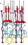 Photoreceptors – the light-sensitive cells in the vertebrate retina – have been extremely well-characterized with regards to their biochemistry, cell biology and physiology. They therefore provide an excellent model for exploring the factors and mechanisms that drive neural progenitors into a differentiated cell fate in the nervous system. Here, and outline the signaling and transcription factors that drive vertebrate photoreceptor development and discuss how these function together in gene regulatory networks to control photoreceptor cell fate specification. See the Review article on p. 3263
A developmental framework for induced pluripotency
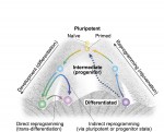 During development, cells transition from a pluripotent to a differentiated state, generating all the different types of cells in the body. Although development is generally considered an irreversible process, it is now possible to reprogram mature cells to pluripotency. Here, and discuss the connections and disparities between differentiation and reprogramming, and assess the degree to which reprogramming can be considered as a simple reversal of development. See the Review article on p. 3274
Featured movie
 (No Ratings Yet)
Get involved
Create an account or log in to post your story on the Node.
Sign up for emails
Subscribe to our mailing lists.